
Prerequisites: What You Need Before You Start

Peptide CoA interpretation starts with documents, not pipettes. Have these five items in hand before you qualify anything:
-
A lot-specific certificate of analysis tied to one exact batch. The document should report identity by MS with observed versus theoretical mass inside a stated tolerance, HPLC purity with the chromatogram and integration table attached, counterion identity, endotoxin in EU/mg with the assay basis named (LAL or rFC), and the analytical method conditions: column, mobile phase and gradient, flow rate, detection wavelength (ICH Q2(R2), 2023-11-30).

-
Raw MS and HPLC data files, not just the summary page, so the record can be reconstructed later.
-
An endotoxin result tied to the sample you will actually test, not to a sibling lot.
-
A handling log covering receipt date, storage temperature, and every thaw, following ALCOA data-integrity principles (MHRA, 2018-03): attributable, legible, contemporaneous, original, accurate.

-
The assay buffer composition, written down, including the final DMSO percentage.
Time, Difficulty, and Assumed Knowledge
Budget two to four hours for a single lot with complete documentation, longer if you need orthogonal confirmation or you are qualifying several lots at once. Difficulty is intermediate. You should already be comfortable reading RP-HPLC chromatograms and integration tables, checking MS mass confirmation against a theoretical value, and interpreting LAL or rFC endotoxin reporting.
Step 1: Verify Peptide Sequence Fidelity Beyond Nominal Purity

By the end of this step you will have confirmed that the peptide’s sequence, not just its chromatographic purity, matches the intended structure. A ≥95% HPLC purity figure describes how much of the peak area is peptide, not which peptide it is, and the two are routinely conflated.
Deletion peptides formed by incomplete coupling are the most abundant sequence-related impurity class in Fmoc SPPS, typically present at the percent-to-subpercent level in crude material and difficult to separate because they differ only subtly from the target (Cellu-Peptide, From Sequence to Vial: Oanpaste peptidesynteze, 2026-07-13). Trifluoroacetate is a different case entirely: it is a counterion rather than a sequence impurity, so it never indicates a wrong sequence, but it does change net peptide content, apparent mass and sometimes retention behaviour, which means it must be corrected for before you calculate assay mass (Towards a Consensus for the Analysis and Exchange of TFA, 2025-08).
Verification checkpoint: the lot’s observed mass matches theoretical within the stated tolerance en you have recorded the counterion content separately from the peptide content.
Reading the MS Confirmation and Counterion Fields
Start with the observed-versus-theoretical mass pair and the tolerance the supplier states. A match inside that window confirms the dominant species; it does not exclude a co-eluting isobaric impurity that happens to ionise at the same m/z.
Counterion content is where peptide CoA interpretation most often goes wrong. In a validated TFA content and exchange study, purified peptide salts carried TFA− up to roughly 35% (w/w), with measured values of 0.333 ± 0.008 mg TFA− per mg peptide salt before exchange, against an expected stoichiometric range of about 22–29% (w/w) depending on charge (Towards a Consensus for the Analysis and Exchange of TFA, 2025-08). If your assay mass comes from nominal peptide weight, that gap lands directly in your molarity.
Foar Tip: Correct for counterion content before calculating assay mass. If the lot needs exchange, the same study found that a single-cycle 10 mM HCl exchange reduced TFA− below the limit of quantitation of all three methods tested (<1% w/w) while purity held above 96% (Towards a Consensus for the Analysis and Exchange of TFA, 2025-08). Note the limitation: routine RP-HPLC and single-stage MS can both miss isobaric impurities, so treat a clean pair of traces as necessary but not sufficient.
When to Request Peptide Mapping or LC-MS/MS
Escalate to an orthogonal method when the sequence carries isomerization-prone or racemization-prone residues, or when the biological readout is unexpectedly strong for the dose you believe you delivered. The rationale is the co-elution problem: closely related sequences that differ by one residue can sit under the same peak, so the chromatogram cannot separate them no matter how the integration is set (Cellu-Peptide, 2026-07-13). Peptide mapping or LC-MS/MS resolves the sequence itself rather than the peak.
Step 2: Control Peptide Aggregation in CNS Assays
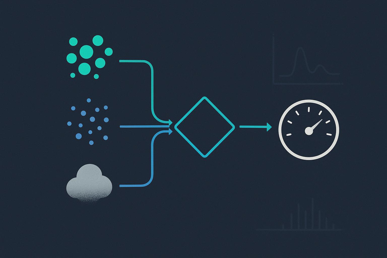
By the end of this step, you will have screened your lot for pre-existing aggregates and cleared them without silently changing the dose you deliver to the assay.
Why Lipophilic Brain-Penetrant Sequences Self-Associate
Aggregation does not require a special motif. IN 2006 study of Aβ42 hydrophobicity and sequence-dependent aggregation showed that generic hydrophobicity is sufficient to drive aggregation: hydrophobic stretches, whether central or C-terminal, can seed fibrils, helix bundles, or disordered aggregates, often passing through dense-liquid or amorphous intermediates before any ordered fibril appears.
That finding matters here because brain-penetrant design pushes sequences in exactly that direction. CNS exposure favors lipophilic character, so the property that makes a peptide useful is the property that makes it aggregate. “Our sequence has no known aggregation motif” is therefore not a reason to skip the screen.
Clearing Aggregates Without Losing Your Dose
Run orthogonal aggregation screening before the assay. Thioflavin T reports amyloid-like fibrils, dynamic light scattering reports particle-size distribution and hydrodynamic radius shifts, and turbidity gives a fast scattering screen. When a signal appears, clear the material by 0.22 µm filtration or centrifugation.
Then re-check concentration. Filtration can remove peptide along with the aggregates, so a filtered stock may no longer match the concentration on the label. No universal rule predicts which sequences will aggregate, which is why the screen is empirical rather than predictive.
Step 3: Confirm Peptide Solubility in CSF-Mimetic Buffer
By the end of this step you will have a documented soluble dose in the actual assay buffer, not a nominal one. Peptide solubility in CSF-mimetic buffer is a measured property of your lot in your matrix, and the gap between the weighed mass and the dissolved mass is where most CNS attribution errors begin.
Prepare the master stock in DMSO, then add it slowly and dropwise into a stirred aqueous phase rather than pipetting it in as a bolus. Vendor guidance on DMSO stock preparation recommends single-use aliquots and a low final DMSO percentage, and treats visible turbidity as a warning sign (Sigma-Aldrich technical guidance; note that this is vendor guidance, and vendors of solvent and reagent products have a commercial interest in the handling practices they recommend). If the solution clouds, the soluble dose is below nominal, whatever the certificate of analysis states.
Buffer Composition and the DMSO Ceiling
Match the buffer to the assay you will actually run, and record the composition with the lot number. Common CSF-mimetic systems include artificial CSF built on a bicarbonate or HEPES base, and phosphate-buffered saline adjusted for pH and ionic strength. Each differs in how it tolerates co-solvent, so the ceiling is a property of the assay, not a fixed number.
For cell-based systems, a final DMSO concentration of 1% or below is the common working limit, and assays that tolerate less should be held to a stricter ceiling. The exact ceiling varies by assay and by source, so establish it for your own readout rather than importing a number from another protocol.
⚠️ Warskôging: Turbidity means the actual soluble dose is below nominal. Do not report the weighed mass as the delivered concentration.
Freeze-Thaw Discipline and Single-Use Aliquoting
Single-use aliquoting is risk reduction, not a hard rule. In a controlled freeze-thaw stress study, measurable particle formation appeared by roughly five cycles in some buffered protein stocks (BSA and β-galactosidase, cycled between −80 °C and 25 °C for 20 minutes each in 100 mM sodium phosphate), while some small molecules showed no loss after eleven cycles (freeze-thaw particle formation in buffered stocks, 2024 pre-proof). That evidence comes from protein stocks rather than peptides, and no universal cycle limit exists, so treat the finding as a reason to aliquot conservatively rather than as a threshold you can rely on.
Step 4: Qualify Endotoxin for Peptide Reagent Quality in Brain-Penetrant NLRP3 Research

By the end of this step you will have an endotoxin result expressed in units tied to the sample you actually tested, with the assay basis and spike recovery documented alongside it. That is the minimum needed to argue the peptide did not contribute to your inflammatory readout.
Why Pass/Fail Endotoxin Reporting Fails a CNS Assay
A “pass” on a certificate of analysis tells you the result fell under a limit someone else set for a different purpose. It does not tell you how much endotoxin is in the vial, so it cannot be converted to a concentration in your well or your animal.
That gap matters because microglia are highly LPS-responsive, and LPS priming followed by a second stimulus such as nigericin readily produces canonical NLRP3 activation. A contaminated CNS peptide can therefore generate a full, convincing inflammasome readout with no contribution from the peptide sequence itself.
The contamination is also hard to spot in your data. LPS priming that does not require new NLRP3 protein remains measurable at short pre-treatment times and persists in Nlrp3−/− macrophages reconstituted with a constitutive promoter. No transcriptional signature appears to flag it.
Unit-Tied Results, Spike Recovery, and Low-Endotoxin Handling
Ask for the result in EU/mg, tied to the tested sample, and for the assay basis behind it. The USP <85> bacterial endotoxins test requires LAL reagent labeled sensitivity not less than 0.15 EU/mL, with limits set per monograph or by the K/M formula and expressed as EU/mL, EU/mg or EU/unit. That compendial floor sits well above what CNS work needs.
Research-grade formats reach lower. Comparative LAL and rFC sensitivity data put quantitation near 0.005 EU/mL for rFC and 0.01 EU/mL for some LAL formats. A commonly used cleanliness target for peptide reagents in cell-based and in vivo CNS work is under 0.1 EU/mL or under 1 EU/mg. Treat that figure as a practitioner target, not a compendial limit. The FDA’s 0.06 EU/mL threshold for CSF-contacting devices is a fluid-contact analogy only, never a peptide specification.
Spike recovery closes the loop. Without it you cannot tell whether the matrix suppressed the assay and produced a falsely low number.
|
Result format |
What it tells you |
What it does not tell you |
Spike recovery needed |
|---|---|---|---|
|
Pass/fail |
The result fell under a stated limit |
The concentration in your assay |
Not applicable |
|
EU/mL |
Concentration in the tested solution |
Dose delivered per mg of peptide |
Yes |
|
EU/mg |
Dose-normalized burden for your weighing |
In-well concentration after dilution |
Yes |
Key Takeaway: An endotoxin result is only usable if it is expressed in units tied to the tested sample, with the assay basis and spike recovery documented.
Step 5: Lock Lot Documentation and Traceability

Tsjinsten By the end of this step you will hold a lot-specific record that can be reconstructed months later, after the person who ran the assay has moved on and the original email thread is gone.
A certificate of analysis that names only a catalog number does not do this. What you need is a document tied to one exact batch, and the fields that make it load-bearing are the ones people skip when they read it quickly.
What a Lot-Specific Analytical Package Should Contain
A lot-specific certificate of analysis should report, for that batch only: MS identity with observed versus theoretical mass and a stated tolerance, HPLC purity with the chromatogram and its integration table, counterion identity, endotoxin in EU/mg with the assay basis named, and the analytical method conditions (column, mobile phase and gradient, flow rate, detection wavelength). Each field answers a different failure question. Mass confirms the sequence; the integration table shows whether the purity figure came from a clean peak or a shoulder; the counterion tells you what else is in the vial; EU/mg ties the endotoxin number to a dose you can actually calculate.
Peptide CoA interpretation is where most labs lose time, because a purity percentage without its chromatogram is not verifiable. A synthesis partner with in-house HPLC/MS and sterility QC can supply a lot-specific analytical package covering these fields, which helps when you need the raw method conditions rather than a summary line.
Resupply Risk Across Multi-Month Studies
A study that runs for months will likely cross lots, and the crossing is usually invisible: the new vial arrives, the label looks the same, and nobody records that the batch identifier changed.
Record at study start the batch identifier, the analytical package revision, and the date each vial entered the freezer. Then apply ALCOA data-integrity principles to the reagent log itself: attributable, legible, contemporaneous, original, accurate, with raw data and calculations preserved alongside the entry. If a mid-study lot change is logged as it happens, a shift in response can be checked against the reagent record instead of being argued about later. Peptide lot-to-lot consistency is not something you can assume from a supplier’s reputation; it is something your own log either documents or does not.
Step 6: Design Labeled and Modified Controls
By the end of this step, you will have a control set that separates label effects from peptide effects, so a fluorescent or affinity readout cannot be mistaken for a biological one.
When a Label Changes the Experiment
A fluorescent or affinity tag is a chemical modification, not a neutral observer. Adding a bulky fluorophore or a biotin handle changes molecular weight, lipophilicity and charge distribution, and those properties govern potency, aqueous solubility and blood-brain barrier behavior. A labeled peptide that partitions differently from the unlabeled reagent can show altered CNS exposure at the same nominal dose, which means a labeled analog is not automatically a valid surrogate for the unlabeled one.
This is the most under-characterized part of the workflow. Most published CNS studies report the label position and little else, so the field has limited comparative Winkel data on how much a given tag shifts potency or brain penetration for a given sequence. Treat label equivalence as an assumption to test, not a property to inherit.
[VISUAL: decision tree, branches on whether the label changes potency, solubility, or BBB behavior, leading to labeled analog, scramble control, inactive analog, or tag-free ortholog]
Scramble, Inactive Analog, and Tag-Free Ortholog Controls
Build the control set around what each reagent rules out, and keep the unlabeled peptide in the experiment wherever the assay can read it directly.
-
Scramble control (same composition, randomized order): rules out sequence-independent effects such as membrane perturbation or nonspecific binding. It does not control for label chemistry, so run it with the same label if the labeled peptide is your test article.
-
Inactive analog (single critical residue substituted): rules out target engagement as the source of the signal. Confirm the substitution actually abolishes activity in your assay rather than assuming it does.
-
Tag-free ortholog or unlabeled parent: rules out the label itself as the cause of the observed effect. Where the readout requires a label, this control can only be run in a parallel label-independent assay.
There is no consensus on the single best control for a given CNS assay. The choice depends on what the readout can detect and on whether the label is load-bearing for the measurement. State your reasoning in the methods rather than citing a convention.
Verify your result: the signal should track the active sequence and disappear with the inactive analog. If the scramble produces the same effect as the test peptide, or if the labeled and tag-free reagents diverge, you have a label or sequence-independent effect, not a target-specific one.
Decision Framework: Reagent Artifact vs. Biological Signal
A CNS readout that moves after peptide treatment has two candidate explanations: the peptide did something, or the reagent did. The distinction matters because the two demand opposite responses. An artifact means the experiment is uninterpretable and the reagent needs requalification; a biological signal means the finding is real and the next step is mechanism work. The table below maps the readout you observed to the reagent check most likely to explain it, and to the result that would clear the reagent.
|
Observed readout |
Peptide synteze Reagent check most likely to explain it |
Result that clears the reagent |
|---|---|---|
|
Cytokine rise with no dose-response |
Endotoxin, re-tested with spike recovery |
Recovery within the assay’s stated window and a result below your assay’s threshold |
|
Steep or erratic dose-response |
Aggregation screen by an orthogonal method |
Monomer-dominant profile at the working concentration |
|
Effect only at the top dose |
Soluble dose in CSF-mimetic buffer |
Full dissolution at the Syntetyske peptiden intended concentration Oer |
|
Activity in the vehicle arm |
Sequence fidelity by orthogonal method |
Confirmed sequence with no dominant deletion species |
|
Signal that tracks the lot, not the dose |
Fresh lot, same protocol |
Reproducible readout across lots |
The Ordered Re-Test Sequence
Run the checks in this order. Each one eliminates a specific class of artifact, so a failure early saves you the sample and time the later steps would consume.
-
Re-check endotoxin with spike recovery. Microglia are highly LPS-responsive, so a priming artifact can mimic a genuine inflammasome effect. Spike recovery tells you whether your buffer or matrix is suppressing the assay, which a pass/fail number alone cannot.
-
Re-screen for aggregates by an orthogonal method. Generic hydrophobicity is sufficient to drive aggregation, and aggregates present as an apparent potency shift rather than as a clean negative.
-
Re-confirm the soluble dose in buffer. Verify the peptide is fully dissolved at the concentration you actually dosed, not at the stock concentration. Peptide produksje
-
Re-verify sequence fidelity by an orthogonal method. Deletion peptides are the dominant sequence-related impurity class, and they can carry partial activity that a nominal purity figure will not reveal.
-
Re-run with a fresh lot. If the readout follows the lot rather than the dose, the reagent is the variable.
When the Signal Survives Every Re-Test
If the effect persists through all five checks, treat it as more likely biological. That is a probabilistic statement, not a certificate. Some artifacts cannot be fully eliminated by any reagent-side check, and orthogonal aggregation screening before the assay narrows the uncertainty without closing it. The framework’s job is to make the residual risk explicit, so that a downstream reader knows which alternative explanations you ruled out and which ones remain open.
Common Mistakes That Produce False Attribution
The most consequential failure mode is not a bad peptide. It is a clean-looking dataset built on a reagent that was never qualified, where a formulation artifact is read as NLRP3 activation and carried forward into the next experiment. Four recurring bench errors produce most of these false attributions.
Treating HPLC Purity as Sequence Proof
The mistake: accepting a 95% of 98% HPLC purity figure as evidence that the peptide is the sequence you ordered.
Why it happens: HPLC separates by hydrophobicity. A deletion sequence, a truncated fragment, or a diastereomer with a single racemized residue can co-elute with the target and still register as a single clean peak. Nominal purity measures separation performance, not structural fidelity.
The fix: require an MS confirmation that matches the theoretical monoisotopic mass, and check the counterion field, because residual trifluoroacetate shifts both mass and biological behavior. Where the assay is sensitive to a single residue change, request peptide mapping or LC-MS/MS fragmentation rather than a mass-only confirmation.
Ignoring the DMSO-to-Buffer Transition
The mistake: diluting a DMSO stock straight into assay buffer and assuming the nominal concentration is the delivered dose.
Why it happens: lipophilic, brain-penetrant sequences have limited aqueous solubility. At the DMSO-to-buffer boundary they can crash out, and the resulting turbidity is often read as acceptable assay noise rather than as lost analyte.
The fix: run the dilution in CSF-mimetic buffer, inspect for turbidity or particulate at the final concentration, and confirm the soluble fraction before you assign a dose. If the solution is not clear, the actual concentration is below nominal regardless of what the calculation says.
Reporting Endotoxin as Pass/Fail
The mistake: recording endotoxin as “passes specification” and moving on.
Why it happens: lot-release certificates are written for release decisions, not for assay design. A pass/fail statement carries no unit-tied value, so it cannot be converted into an in-assay concentration.
The fix: request the result in EU/mg or EU/mL, tied to the tested sample, with spike recovery documented. Then calculate what that mass of peptide contributes at your working concentration. A lot that passes release can still deliver a confounding TLR4 signal at a high micromolar dose.
Reusing a Freeze-Thawed Stock
The mistake: pulling the same aliquot out of the freezer for each replicate across a multi-week study.
Why it happens: freeze-thaw cycling is treated as harmless for a “stable” peptide, and single-use aliquoting looks like unnecessary work.
The fix: aliquot at first reconstitution and discard after one use. Particle formation accumulates across cycles, and the aggregate fraction is exactly the species most likely to drive an inflammasome readout. Note the scope here: the freeze-thaw literature is largely built on protein stocks, not peptides, and no universal cycle limit applies. Treat the number of cycles as a variable you control and document, not a threshold you can look up.
Verify Your Result: Qualification Checkpoints
Qualification is finished when you can hand a reviewer one lot-specific record containing all six items below. Each is verifiable against a document, not a memory of what the vendor said on the phone.
-
Identity: an MS-confirmed molecular mass that matches the sequence you ordered, plus an orthogonal check (peptide mapping or LC-MS/MS) wherever the sequence or modification warrants one.
-
Aggregation: a cleared screen result at your working concentration, with the method and the concentration recorded.
-
Solubility: a documented soluble dose in your CSF-mimetic buffer, including the final DMSO percentage.
-
Endotoxin: a unit-tied result (EU/mg or EU/mL) with the spike recovery that validates the assay in your matrix.
-
Controls: a defined set of scramble, inactive analog, and tag-free ortholog reagents, each qualified to the same standard as the test peptide.
-
Traceability: lot number, release date, and the analytical package that ties every field above to that lot.
The stretch goal is to extend this from a per-lot record into a study-level reagent qualification log: one row per lot, one column per checkpoint, so a multi-month study can show at a glance that every batch entered the assay under the same qualified conditions.
[VISUAL: before-after, left panel shows the Step 1 state (a CoA with a purity percentage and nothing else); right panel shows the completed record with identity, aggregation, solubility, endotoxine, control, and traceability fields filled in]
If you want that record supplied rather than assembled, request a lot-specific analytical package for the sequence you are running, or open a technical discussion on custom or modified peptide requirements. MOL Changes publishes as a peptide vendor and has a commercial interest in peptide quality standards; the checkpoints above are the same ones we would expect a reviewer to apply to us.
Beoardiele troch [Name], Ph.D., Analytical Chemistry.
Getting peptide reagent quality for brain-penetrant NLRP3 research to a defensible standard is a documentation exercise as much as an analytical one: the assays tell you what is in the vial, and the record is what lets anyone else confirm it.
Faak stelde fragen
How long does peptide reagent qualification take?
Plan for two to four weeks from lot receipt to a signed qualification record. Three variables move that window: whether orthogonal sequence methods such as peptide mapping or LC-MS/MS are needed, whether endotoxin testing runs in-house or goes to a contract lab, and whether a fresh lot has to be sourced. If the lot arrives with a complete analytical package and in-house endotoxin capacity, the work compresses toward the lower end.
Can I substitute a small-molecule NLRP3 inhibitor as a positive control?
Only as a pharmacological control, not as a reagent qualification substitute. The primary literature centers small-molecule NLRP3 inhibitors, so they are well characterized for pathway engagement, but they tell you nothing about whether your peptide reagent is behaving. Run both: the small molecule confirms the assay responds, and the peptide lot confirms your reagent is clean.
What should I do if my peptide fails the aggregation screen?
Re-filter or centrifuge the stock, then re-check concentration before assuming the lot is unusable. IN 0.22 µm filtration step clears pre-existing aggregates, but you must re-measure concentration afterward because material is lost on the filter. If the sample still aggregates, adjust the DMSO-to-buffer transition rate or request a different lot.
Is a labeled peptide an acceptable substitute for the unlabeled reagent?
It depends on whether the label changes potency, solubility, or blood-brain barrier behavior. A fluorophore or tag can shift all three, so characterize the labeled analog separately rather than assuming equivalence. Use the labeled peptide for detection and the unlabeled reagent for the primary readout.
How do I qualify a peptide for a study that runs longer than one lot?
Record the lot number at study start, then qualify every new lot against the same checkpoints you used initially. Bridge lots by running a side-by-side comparison on the same assay plate before switching. Document any mid-study lot change, including the qualification data, so the switch is traceable in the record.