
Step 1: Define the Assay Objective and the Bottleneck It Creates
Before you compare labels or linkers, name the one thing your assay cannot currently do. That single failure point, not the modification menu, decides which options are worth your time.
Most peptide modification selection by assay objective reduces to four bottlenecks. If the readout is weak or unquantifiable, you have a detection problem. If the peptide will not stay where the assay needs it, you have a capture or immobilization problem. If activity disappears before the readout window closes, you have a half-life or protease resistance problem. If the peptide behaves differently from the native sequence at its termini, you have a native-like terminal behaviour problem.

The distinction matters because each bottleneck points at a different family of chemistry. Bachem’s overview of peptide modification frames modification choice as a function of the role the peptide must play: reporter groups for detection and quantification, affinity handles for capture and immobilization, stabilization features for half-life and protease resistance, and conjugates for delivery. Peptide labeling and conjugate design is therefore a downstream decision, not a starting point.
Klíčové s sebou: Sort every candidate modification against one of four bottlenecks: detection, capture/immobilization, half-life or protease resistance, or native-like terminal behaviour. That axis carries through the rest of this guide.
Step 2: Map the Objective to a Modification Class
Peptide modification selection by assay objective works best as a mapping exercise: name the bottleneck, then read across to the class that addresses it and the specification field that class forces you to define. The modification classes available to researchers span terminal changes, side-chain chemistry, backbone edits, labels, conjugates and conformational constraints (Bachem peptide modification overview, retrieved 2026-03-15).
|
Assay objective / bottleneck |
Služby Modification class |
Specification field it forces |
|---|---|---|
|
Prevent N- or C-terminal degradation |
N-terminal acetylation, C-terminal amidation, pyroglutamate formation |
Terminal group, stated explicitly on the order |
|
Detect or capture the peptide |
Biotinylation; fluorescent labels (FITC, FAM, TAMRA, Cy3/Cy5/Cy7) |
Label identity and conjugation site |
|
Raise immunogenicity for antibody production |
Carrier-protein conjugation (KLH, BSA, OVA) |
Carrier, coupling chemistry, peptide-to-carrier ratio |
|
Extend circulation or reduce renal clearance |
PEGylation, polymer conjugation |
Polymer molecular weight and attachment point |
|
Constrain conformation for binding studies |
Cyclization (head-to-tail, side-chain, disulfide), hydrocarbon stapling |
Bridge positions and cyclization type |
|
Block oxidation of a methionine |
Methionine replacement with norleucine |
Substitution position and its effect on activity |
|
Resist protease digestion |
D-aminokyseliny, N-methylated and non-proteinogenic residues |
Which residues are modified, and where |
Syntéza peptidů Two rows deserve emphasis. N-terminal acetylation and C-terminal amidation are the terminal changes nature uses to stabilize peptides, which is why they appear in so many native protein segments (Bachem peptide modification overview, retrieved 2026-03-15). And backbone modifications, depsipeptides and C-terminal esters or thioesters change the chemistry of the chain itself, so they interact with every other choice you make downstream.
Step 3: Decide Where the Modification Goes
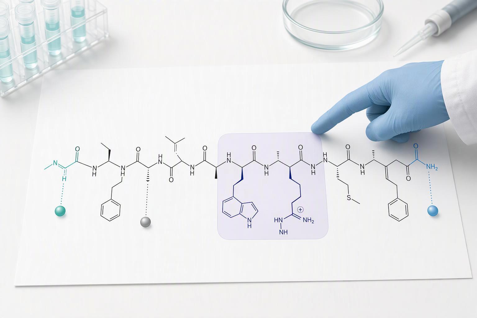
Placement is a separate decision from modification class, and it is where most readouts are lost. The rule is simple: label where it least interferes with binding, and use a spacer when surface access matters. Merck’s guidance on peptide design sets out N-terminal, internal and C-terminal placement options, with the terminus-first logic holding unless a defined residue has to be addressed directly.
The failure mode is a masked epitope. If the active motif sits within two residues of the N-terminus, an N-terminal label can sit on top of it, and the assay reads a weaker interaction than the peptide actually supports. Move the label to the opposite terminus, or to an internal position outside the motif, and the same sequence recovers its signal. When the payload is bulky or has to be presented away from the epitope, a spacer restores access: Bachem’s guide to biotinylated peptides notes that the typical optimal spacer length between biotin and peptide is 6 na 12 atoms, because short spacers cause steric hindrance while longer spacers raise cost. That trade-off is the whole placement decision in one line.
Placement also interacts with chemistry inside the same class. In a 2024 comparison published in the International Journal of Molecular Sciences, lactam-stapled analogues reached 2.9 h and 5.0 h in rat plasma, while the corresponding hydrocarbon staples reached 4.0 h and 6.8 h. Same stabilization intent, different measured outcome.
Step 4: Lock the Specification Fields Before You Order
By the end of this step you have a specification sheet that a supplier can quote against without guessing. The Clinical Chemistry recommendations on peptide antibody characterization set out the specification fields to settle before ordering: sequence and modification identity, attachment site, degree of labeling or stoichiometry, counterion, salt form, solubility and solvent, purity grade, and the characterization evidence you require on the certificate of analysis.
Purity grade is the field buyers most often leave implicit. Supplier guidance from Bachem’s guide to biotinylated peptides states that the 95% purity expectation applies to most peptide applications, where the goal is reducing background and off-target effects. Treat that as supplier guidance with vendor bias rather than a measured distribution, and match the grade to what the assay can tolerate.
Counterion deserves its own line. Work gathered on residual TFA and counterion exchange reports residual TFA in TFA-salt lyophilized peptides commonly at roughly 10 na 30% of mass, falling below 1% after acetate exchange, with ion chromatography as the gold-standard method. An aggregator range up to 45% circulates online but is an unconfirmed upper estimate, so do not plan around it.
|
Specification Shop field |
What to state on the order |
|---|---|
|
Sequence and modification identity |
Exact modification, not a category label O |
|
Attachment Syntetické peptidy site |
Residue number or terminus |
|
Degree of labeling or stoichiometry Výroba peptidů |
Target ratio, with tolerance |
|
Counterion and salt form |
TFA or acetate, stated explicitly |
|
Solubility and solvent |
Working solvent and target concentration |
|
Purity grade |
Percentage, tied to assay background tolerance |
|
Characterization evidence |
Tests required on the CoA |
For custom modified peptide synthesis specifications, this table is the document to send.
Step 5: Verify the Delivered Material Against the Modification, Not Just the Purity
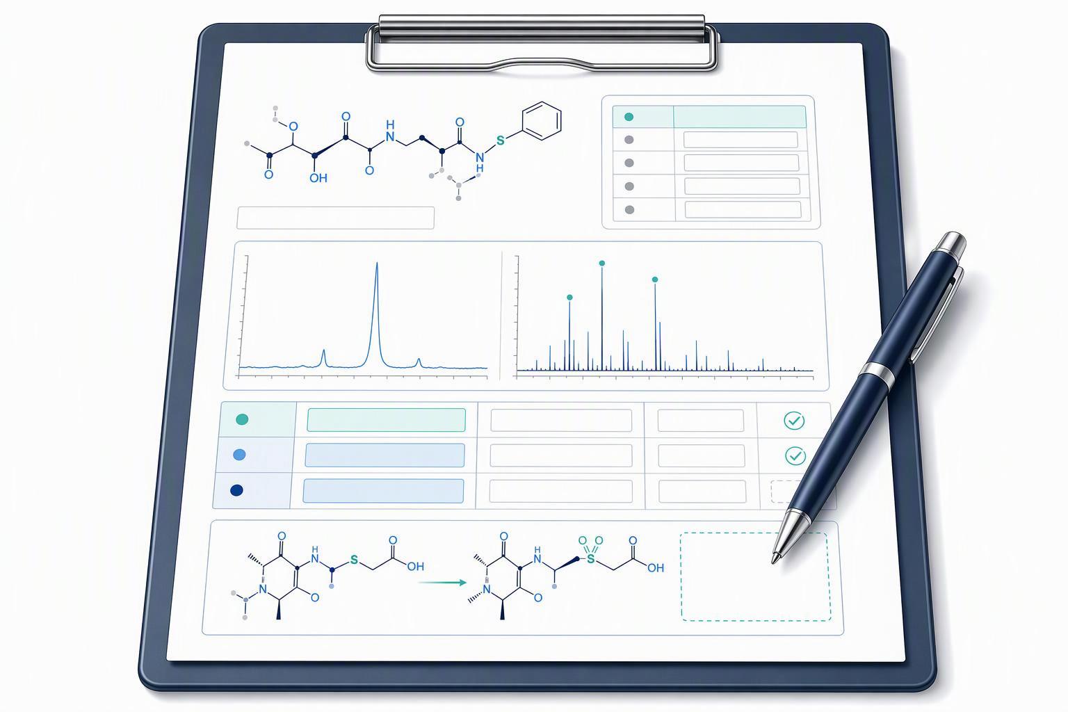
A certificate of analysis that reports 98% purity and a matching mass has confirmed two things: the material is largely one peptide, and its mass is the mass you ordered. It has not confirmed that the modification sits where you specified. Mass spectrometry is what closes that gap. It confirms the added mass, and with MS/MS fragmentation it can establish the site of modification, while HPLC co-elution is supporting identity evidence only and does not by itself prove the modification (PMC, 2024). For a dye, UV/Vis or fluorescence quantifies how much label is present but says nothing about where it attached. For biotin, streptavidin capture is the functional orthogonal check that the label still binds.
HPLC with mass spectrometry is the standard verification pairing for purity, sequence and successful biotin incorporation (Bachem). Read the mass accuracy the same way: on modern high-resolution LC-MS, peptide identity confirmation typically sits at ±5 ppm, with 5 na 10 ppm as the practical search window and anything above about 20 ppm treated as suspicious (Peptidepedia, 2025). Treat that as a practice range, not a specification you can hold a supplier to.
Klíčové s sebou: Match the evidence to the modification class. MS proves added mass; MS/MS proves site; HPLC proves purity and supports identity; UV/Vis or fluorescence proves label quantity, not position; a functional assay such as streptavidin capture proves the conjugate still works. A CoA carrying only purity and mass leaves placement unverified.
Step 6: Match the Stabilization Strategy to the Required Half-Life

Stabilization is a quantitative decision: the target half-life you need sets the strategy, and the strategy sets the cost and the analytical burden. In human serum, cyclization moved PRP-1 from a linear half-life of 0.41 ± 0.02 h to 7.7 ± 0.4 h, roughly 19-fold, and PRP-3 from 0.21 ± 0.02 h to 28 ± 5 h, roughly 133-fold (Serum Stable Natural Peptides Designed by mRNA Display, 2014). Both figures come from one upstream study, so treat the direction as reliable and the exact fold-change as study-specific.
PEGylation behaves differently: it tunes exposure rather than locking the backbone. In mouse serum, temporarily PEGylated Onc72 released at 8 h with a 5 kDa chain and 14 h with a 20 kDa chain, and elimination half-life in female CD-1 mice at 4.34 µmol/kg subcutaneous was 66 min at 5 kDa and about 5.5 h at 20 kDa, against 43 min for free Onc72 (PEG chain length and elimination half-life in mice, 2023). These are mouse pharmacokinetics, not human half-lives.
Choosing a peptide stabilization modification without a target half-life is the failure mode here: you pay for cyclization or a large PEG chain and still miss the window your assay needs.
Common Mistakes to Avoid
Most failed custom peptide orders trace back to a specification gap, not a synthesis error. The documented failure modes are masked epitopes, mixed or heterogeneous labeling including positional isomers, altered charge and solubility, affinity loss from bulky tags, and aggregation from lipidation (PMC review of peptide design and formulation constraints, 2020-10-28).
Specifying a modification without naming its attachment site. A request for “biotinylated peptide” leaves the position to the chemist, and the result can be a positional isomer mixture rather than the single species your assay assumes. Name the residue and its number.
Treating purity as proof of the modification. An HPLC purity figure describes how much material eluted as one peak, not whether the label is present or where it sits. Mass spectrometry confirms identity; only site-specific analysis confirms placement.
Ignoring counterion effects on solubility. The same sequence can behave differently in assay buffer depending on the counterion it was supplied with. State your buffer and solubility expectation before ordering.
Placing a bulky tag over a binding-critical motif. Large labels can sterically block the interface your assay depends on, which surfaces as affinity loss rather than a failed QC result.
Choosing a stabilization strategy without a target half-life. Cyclization and stapling change stability, but without a stated half-life requirement you cannot tell whether the modification was sufficient or excessive.
Warning: An unnamed attachment site cannot be corrected after synthesis, and the resulting isomer mixture may still pass purity and mass checks while invalidating your binding data.
When you select a labeling site, avoid oxidation-prone residues: Cys, Met, His, Trp and Tyr are the residues most susceptible to oxidation at a modification site, and deamidation hotspots follow the NG, NS, NN, NT and NH motifs (PMC review of protein oxidation and post-translational modification chemistry, 2015-09-21). If phosphorylation must survive, keep Ser, Thr and Tyr clear of the modification position, and avoid the N-X-S/T sequon when N-linked glycosylation has to be preserved.
What Success Looks Like and What Comes Next
A correct run of this framework produces two artifacts. The first is a specification sheet in which every field traces back to a named assay objective: the modification class, its position, the spacer or handle, the purity grade, and the confirmation method you will accept. The second is a verification plan whose evidence matches the modification rather than the peptide alone, so the certificate of analysis answers the modification-specific question instead of only the purity question.
The standards anchor for that verification plan is the ICH Q2(R2) validation framework, adopted at Step 4 alongside Q14 on 1 November 2023 and issued as FDA final guidance with content current as of 6 March 2024. It sets out general principles for analytical procedure validation, including the analytical use of spectroscopic data, which is the same reasoning you apply when deciding what evidence a labeled or conjugated batch needs.
The stretch goal is lot-to-lot consistency. Once peptide modification selection by assay objective is documented for one order, the same specification sheet becomes the acceptance template for repeat batches, and drift shows up as a failed field rather than a failed experiment.
If your next project involves a modification outside your current experience, a technical conversation before ordering is usually cheaper than a repeat synthesis.
Disclosure: MOL Changes has a commercial interest in peptide quality standards and offers custom modified peptide synthesis. This article is written for research use; it is not clinical or diagnostic guidance.
Často kladené otázky
Can a modification be added after synthesis instead of during it?
Some can, most cannot. N-terminal acetylation, C-terminal amidation, and PEGylation are typically installed as the final synthetic steps, while side-chain and non-natural residue work has to be built into the chain as it grows. Post-synthesis labeling is practical when the target is a single reactive group on an otherwise finished peptide. If the modification must sit at a defined internal position, plan it into the sequence before the first coupling.
Fluorescent label or biotin tag when the assay needs both detection and capture?
Use both, but decide which one carries the quantitative signal. Biotin gives you streptavidin capture and pull-down; a fluorophore gives you direct readout. When both sit on the same peptide, check that the label does not sit near the biotin site, because a bulky fluorophore can sterically hinder streptavidin binding. A short spacer between the two usually resolves it.
What if the required half-life exceeds what cyclization alone delivers?
Cyclization mainly restricts conformational flexibility and can improve resistance to exopeptidases, but it does not stop every degradation route. Where cyclization is not enough, the literature strategies are stapling, D-amino acid substitution at scissile positions, and PEGylation to raise the effective size. Which one applies depends on which protease is doing the damage, so identify the degradation route first rather than stacking modifications.
Is a lower purity grade acceptable for a screening assay?
Often yes, with a caveat. Crude or 70-80% material is workable for early screening where the readout is a relative ranking, provided you know what the impurities are. For anything quantitative, or for cell-based work where residual reagents can confound the result, use higher purity. If the assay touches endotoxin-sensitive systems, note that release testing for GMP-grade material includes endotoxin not more than 10 EU/mg by USP <85> and sterility by USP <71> (GMP peptide release specifications), and the wider USP endotoxin limit framework sets 5 EU/kg/hour for most parenteral routes and 0.2 EU/kg/hour for intrathecal.
Conclusion
Choosing peptide modifications by assay objective comes down to running one sequence: name the objective and the bottleneck it creates, map that to a modification class, decide where the modification sits, lock the specification fields before ordering, verify the delivered material against the modification itself, and match the stabilization strategy to the half-life the assay actually needs. The framework exists to prevent one specific failure: a peptide that clears every purity check on the certificate of analysis, arrives with a clean HPLC trace and a matching mass, and still fails the assay because the modification was never independently verified. Purity and identity are not the same question as modification integrity, and only the second one tells you whether the label, the conjugate, or the terminal change is where you intended it to be. Before your next custom modified peptide synthesis order, take the specification checklist from Step 4 and confirm each field against the assay objective you wrote down in Step 1.