Luminex Cytokine Panels hauv Organoid Studies: Peptide Reagent Interference and Experimental Controls
Peptide Synthesis Three-dimensional (3D) organoid models—tshwj xeeb cerebral, neuroimmune, thiab intestinal human organoids—tau hloov pauv translational disease modeling. Los ntawm kev rov piav txog tissue architecture thiab cell-type diversity, organoid cultures allow researchers to measure localized cytokine and chemokine secretion in response to therapeutic agents, viral mimics, or synthetic peptide agonists. Bead-based multiplex immunoassays, such as Luminex xMAP technology, represent the gold standard for profiling these complex supernatants due to their high throughput, broad dynamic range, and minimal sample volume requirements (25–50 μL).
However, quantifying neuroimmune mediators in organoid conditioned media presents unique analytical challenges. Synthetic peptides introduced into cultures as receptor ligands, therapeutic candidates, or disease aggregators (e.g., beta-amyloid, alpha-synuclein, or viral epitopes) frequently generate severe artifactual readouts. When combined with viscous extracellular matrix (ECM) hydrogels like Matrigel or Geltrex, these reagents trigger non-specific bead cross-reactivity, reporter signal quenching, optical bleed-through, and prozone effects. Without rigorous experimental controls, these artifacts produce false-positive inflammation signatures or mask true biological responses.
This technical guide outlines the biophysical mechanisms driving synthetic peptide and label interference in Luminex assays, presents quantitative spike-and-recovery control protocols, and details sample preparation strategies to ensure robust, publication-grade multiplex cytokine data.
Biophysical Mechanisms of Synthetic Peptide and Label Interference
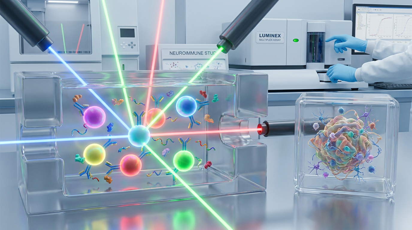
Luminex xMAP technology relies on polystyrene or magnetic microspheres internally dyed with precise ratios of red and infrared fluorophores. A red laser or LED (635 nm) identifies the specific bead region (analyte identity), while a green laser or LED (532 nm) excites Streptavidin-Phycoerythrin (SAPE) bound to biotinylated detection antibodies to quantify the target analyte (Mean Fluorescence Intensity, MFI). Synthetic peptides thiab lawv cov kev hloov tshuaj lom neeg sib cuam tshuam nrog txhua theem ntawm txoj kev optical thiab immunochemical no.
Non-Specific Bead Adsorption via Hydrophobic and Electrostatic Patches
Cov synthetic peptides uas tsis tau hloov thiab hloov kho feem ntau muaj amphipathic, tsis nyiam dej heev, lossis polycationic sequences domains. Polycationic sequences uas muaj arginine-rich lossis TAT-like cell-penetrating motifs txuas electrostatic rau saum npoo carboxylated ntawm magnetic beads (MagPlex). Zoo ib yam, hydrophobic peptide clusters (muaj leucine, isoleucine, lossis tryptophan) adsorb tsis tshwj xeeb rau polystyrene microspheres.
Qhov kev txuas tsis tshwj xeeb no tsim ib txheej protein ntawm lub pob uas ntes tau cov tshuaj tiv thaiv kab mob lossis SAPE reporter molecules ywj pheej ntawm qhov cytokine hom phiaj. Consequently, cov kws tshawb fawb pom MFI nce siab thoob plaws ntau txoj kev cytokine uas tsis sib txuas, mimicking broad neuroimmune activation.
High-Dose Prozone (Hook) Effects in Peptide Stimulation
In functional organoid assays, synthetic peptides are frequently applied at micromolar concentrations to stimulate receptors or model protein aggregation. When conditioned supernatants containing high residual peptide concentrations or peptide-induced cytokine levels exceeding 50,000 pg/mL are assayed without adequate dilution, the prozone (high-dose hook) effect occurs.
Unbound analyte or cross-reactive peptide fragments saturate both the bead-bound capture antibodies and the soluble biotinylated detection antibodies simultaneously. This prevents the formation of the classic “sandwich” complex (Capture Ab – Analyte – Detection Ab-Biotin). Instead, separate capture-analyte and detection-analyte binary complexes form in solution, causing a sharp, kev poob qis paradoxical hauv MFI signal txawm tias muaj concentration tshuaj ntsuam siab heev.
Optical Crosstalk and Bead Misclassification from Fluorescent Labels
Fluorophore-tagged peptides (e.g., FITC, FAM, Cy5, lossis Alexa Fluor 647) tau siv dav hauv kev kawm organoid uptake thiab receptor-binding. Cov fluorophores seem nyob hauv cov khoom kho mob ua rau muaj kev cuam tshuam loj ntawm kev pom Luminex:
- Reporter Channel Crosstalk: Fluorophores xws li FITC lossis FAM muaj spectra dav (peak emission ~520 nm) uas sib tshuam nrog SAPE reporter channel (emission peak ~575 nm). Fluorophores uas tsis tau txuas lossis tsis tau adsorbed tshwj xeeb tsim fluorescence cuav hauv 532 nm laser excitation, nce baseline MFI thiab ua rau muaj false positive.
- Bead Misclassification: Far-red fluorophores zoo li Cy5 lossis Alexa Fluor 647 tso teeb hauv 660–680 nm qhov rai, sib tshuam ncaj qha nrog internal bead-classification red laser optics (635 nm excitation). This spectral bleed-through shifts the apparent spectral signature of the microspheres, causing the Luminex instrument to misclassify beads into wrong analyte regions or reject them as “unassigned beads,” resulting in low bead counts and missing data points.
Depletion of SAPE Reporter via Biotin-Tagged Reagents
Biotinylated synthetic peptides are standard tools for affinity pull-downs and pulse-chase labeling in organoid workflows. However, free biotinylated peptides present in culture supernatants present a critical mechanism of interference: direct competition for the SAPE reporter conjugate.
During the secondary incubation step, free biotinylated peptides bind with sub-picomolar affinity to Streptavidin-Phycoerythrin in solution. This exhausts the available SAPE pool, preventing reporter conjugation to the biotinylated detection antibodies immobilized on target cytokine beads. Rau qhov no, cov cim cytokine tiag tiag raug txo qis heev, ua rau muaj kev nyeem tsis raug.
Qhov tseem ceeb: Fluorophores tso tawm ze 575 nm inflate tus reporter MFI, thaum cov cim liab heev ua rau kev cuam tshuam 635 nm bead-classification channel. Cov peptides biotinylated dawb sib tw ncaj qha rau SAPE, txwv tsis pub cytokine tiag tiag txog qhov twg 90%.
ECM Matrix Effects: Addressing Matrigel and Organoid Media Suppression
Ib puag ncig kev loj hlob ntawm 3D organoids nyob ntawm cov hydrogels extracellular matrix nyuaj heev (Matrigel, Geltrex, lossis recombinant collagen matrices) thiab cov khoom tshwj xeeb tsis muaj serum nrog cov khoom loj hlob, B27, thiab N2. Cov khoom ntxiv no tsim kev cuam tshuam matrix loj uas txawv ntawm kev loj hlob ib txheej ib txwm.
Organoid Supernatant Matrix Challenge
Soluble ECM Hydrogels (Laminin, Collagen IV, HSPGs)
Nce viscosity ntawm cov qauv
Npog microspheres → thaiv kev ntes antibodies
Media Additives & Growth Factors (BDNF, GDNF, FGF-2)
Cross-reacts with structural cytokine homology domains
Residual Cell Debris & Microparticles
Induces bead aggregation & capillary clogging in fluidics
Soluble basement membrane proteins, particularly laminin and collagen IV fragments, increase supernatant viscosity and coat microspheres in a non-specific gel layer. This physical barrier restricts antibody-antigen mass transport, causing non-linear signal suppression. u, high concentrations of recombinant growth factors added to neural organoid media can cross-react with specific cytokine capture antibodies due to structural domain homology, generating false baseline signals.
Researchers evaluating complex matrices can reference peer-reviewed evaluations of multiplex matrix suppression to understand how standard assay diluent formulations often fail to fully reverse matrix-induced inhibition without customized surfactant and blocking modifications.
Quantitative Control Protocols and Assay Validation Formulas
To distinguish authentic biological cytokine responses from synthetic peptide and matrix artifacts, researchers should structure their validation routines around international bioanalytical guidelines, such as ICH Q2(R1) and FDA Bioanalytical Method Validation standards. Prior to analyzing experimental organoid samples, laboratories must execute three quantitative validation protocols to benchmark accuracy, precision, and matrix tolerance.
1. Spike-and-Recovery Assessment
Spike-and-recovery experiments evaluate whether the sample matrix or synthetic peptide reagent alters the expected signal of a known analyte concentration. Known quantities of recombinant cytokine standards are spiked into three parallel vehicles:
- Standard Assay Diluent (C diluent)
- Unstimulated Organoid Conditioned Media (C matrix)
- Organoid Conditioned Media + Synthetic Peptide Reagent (C matrix+peptide)
The Percent Spike Recovery (R%) is calculated as:
R% = C spiked sample – C unspiked sample{C spiked diluent} × 100%
- Acceptance Criterion: 80% ≤ R% ≤ 120%.
- Interpretation: Recovery below 80% indicates matrix suppression or SAPE depletion by biotinylated reagents. Recovery above 120% signals additive fluorescence crosstalk or non-specific binding.
2. Matrix Effect Suppression Percentage
To isolate matrix-induced quenching from optical additive noise, calculate the Matrix Effect Percentage (ME%):
ME% = ( 1 – MFI_{spiked matrix} – MFI_{unspiked matrix}{MFI_{spiked diluent} – MFI_{blank diluent}} ) × 100%
A value of ME% > 15% mandates sample dilution or diluent modification with non-ionic surfactants.
3. Serial Dilution Parallelism Linearity
Dilution parallelism confirms that the analyte response behaves proportionally across serial dilutions, establishing that matrix or peptide interference is eliminated at a specific working dilution.
Organoid supernatants are diluted 1:2, 1:4, 1:8, thiab 1:16 in optimized assay diluent. Corrected concentrations are calculated by multiplying measured values by the dilution factor:
Corrected Concentration = Measured Concentration × Dilution Factor
- Acceptance Criterion: Observed concentrations across consecutive dilutions must maintain recovery within 85–115% of the initial diluted value, with a linear regression coefficient R² ≥ 0.98. Non-linear increases in corrected concentration upon dilution indicate relief from prozone effects or matrix suppression.
Pro Tip: Always run a “peptide-only” control well containing the synthetic peptide reagent in assay diluent without organoid supernatant across the entire multiplex panel. Any MFI above background in this well indicates direct peptide-bead cross-reactivity or label crosstalk.
Empirical Case Snapshot: Neuroimmune Organoid IL-6 and TNF-α Spike Recovery
To illustrate the practical impact of these controls, Table 1 displays representative benchmark data from human neuroimmune organoid conditioned media evaluating IL-6 and TNF-α recovery before and after two-stage centrifugation and 1:8 diluent optimization (0.05% Tween-20 + 1% BSA).
| Condition / Analyte | Unclarified Supernatant (1:2 Dilution) | Clarified Supernatant + Optimized Diluent (1:8 Dilution) | Validation Outcome | ||||
|---|---|---|---|---|---|---|---|
| IL-6 Spike Recovery (R%) | 58.4% (Severe Matrix Suppression) | 97.2% (Optimal Recovery) | Pass (80–120% target) | ||||
| TNF-α Spike Recovery (R%) | 134.1% (Non-Specific Cross-Talk) | 102.5% (Baseline Normalized) | Parallelism Linearity (R²) White Label Peptides Manufacturer
tr> |
Parallelism Linearity (R²) | 0.81 (Non-linear prozone shift) | 0.992 (High Linearity) | Pass (R² ≥ 0.98) |
Cov kev ntsuas no qhia tau tias kev ntxuav ceev siab nrog kev ntxiv surfactant tsis ionic rov qab tau qhov tseeb hauv kev lees paub bioanalytical.
Practical Sample Preparation and Mitigation Protocols
Kev siv pre-treatment thiab assay buffer optimization nruj yuav tshem tawm feem ntau ntawm peptide thiab ECM hydrogel artifacts.
1. Ob-Theem Centrifugation thiab ECM Clarification
Organoid supernatants yuav tsum dhau ob kauj ruam kev ntxuav sai tom qab sau:
- Low-Speed Centrifugation: Spin supernatants ntawm 1,000 × g rau 10 feeb ntawm 4°C kom pellet cov hlwb thiab cov khoom organoid loj.
- High-Speed ECM Clarification: Hloov sab saud 80% ntawm supernatant mus rau raj tshiab thiab centrifuge ntawm 16,000 × g rau 15 feeb ntawm 4°C, lossis dhau los ntawm low-binding 0.22\ μm PVDF membrane filter. Kauj ruam no tshem tawm cov matrigel micro-aggregates uas tsis soluble uas ua rau capillary clogging thiab bead clumping hauv Luminex fluidics.
2. Diluent Optimization nrog Non-Ionic Surfactants thiab Blockers
Cov tshuaj diluents assay qauv yuav tsum tau ntxiv kom txo kev sib cuam tshuam ionic thiab hydrophobic ntawm synthetic peptides thiab microspheres:
- Non-Ionic Surfactants: Add 0.05%\ v/v Tween-20 lossis Triton X-100 kom txo hydrophobic adsorption yam tsis denatured capture antibodies.
- Carrier Blockers: Muaj 1%\ w/v Bovine Serum Albumin (BSA) los yog 0.5%\ nrog casein nyob rau cov chaw txuas carboxyl tsis tshwj xeeb ntawm cov pob zeb.
- Heterophilic Blockers: Add 100\ μg/mL cov tshuaj tiv thaiv antibody uas tsis muaj kev tiv thaiv kab mob lossis cov tshuaj tiv thaiv heterophilic uas muaj muag hauv khw (HBT) txhawm rau tshem tawm kev cuam tshuam ntawm kev sib txuas.
3. Pre-Incubation Free-Label Depletion
Rau kev tshawb fawb siv cov peptides uas muaj biotinylated lossis fluorophore-tagged peptides, cov cim npe uas tsis tau teb yuav tsum raug tshem tawm p
Free Biotin Depletion: Pass biotinylated peptide stocks through a monomeric avidin column Peptide Synthesis or perform size-exclusion chromatography (SEC) to ensure >99.5% purity of conjugate over free biotin.
on chromatography (SEC) to ensure >99.5% purity of conjugate over free biotin.
- Free Dye Removal: Utilize reverse-phase solid-phase extraction (SPE) los yog 1 kDa spin-dialysis to eliminate free FITC/Cy5 fluorophores.
For detailed protocols on peptide stimulation kinetics and avoiding high-dose saturation, consult literature on peptide stimulation optimization in Luminex workflows.
Peptide Synthesis When aberrant MFI signals or high background values occur during organoid cytokine profiling, ua raws li txoj kev kuaj mob muaj qauv:
r during organoid cytokine profiling, ua raws li txoj kev kuaj mob muaj qauv:
[Aberrant Luminex MFI Signal Detected]
   │
   ├── Question 1: Is MFI elevated in blank wells containing synthetic peptide alone?
   │     ├── YES ──► Optical crosstalk or non-specific bead adsorption.
   │     │             Action: Switch to non-overlapping fluorophore; add 0.05% Tween-20.
   │     └── NO  ──► Proceed to Question 2.
   │
   ├── Question 2: Does serial dilution (1:2 to 1:16) yield non-linear increases in calculated concentration?
   │     ├── YES ──► Prozone hook effect or ECM matrix suppression present.
   │     │             Action: Assay samples at higher working dilution (e.g., 1:8).
   │     └── NO  ──► Proceed to Question 3.
   │
   ├── Question 3: Is Spike Recovery (R%) in conditioned media < 80%?
   │     ├── YES ──► SAPE depletion by free biotin or physical ECM coating.
   │     │             Action: Perform streptavidin pre-clearing or 0.22 μm PVDF filtration.
   │     └── NO  ──► Assay is validated. Signal represents authentic cytokine response.
Reviewing technical specifications on xMAP bead classification optics and SAPE detection chemistry helps researchers select compatible reporter systems when designing multiplex panels.
Peptide Quality Standards for Assay-Grade Controls
The validity of any multiplex immunoassay relies fundamentally on the purity and structural integrity of the synthetic peptides and controls utilized. Standard research-grade peptides frequently contain unreacted coupling reagents, truncated sequences, free fluorophores, and trace endotoxins that corrupt organoid culture responses and immunoassay readouts.
| Standard Research-Grade Peptides Assay-Grade Synthetic Controls | Detail |
|---|---|
| Kev huv si | 80-90% (Truncations present) ► Purity: >98% by C18 RP-HPLC |
| Environment Pi Of Peptide Supplier | Up to 5% free fluorophore ► Free Label: <0.1% via ESI-MS |
| Endotoxin | Unspecified (>10 EU/mg) ► Endotoxin: Certified <0.01 EU/mg |
| Environment | Standard laboratory ► Environment: Class 100 Sterile Cleanroom |
To prevent artifactual cytokine induction, R&D scientists should source control peptides manufactured under stringent Quality Assurance parameters:
- Purity Verification: Minimum >95–98% purity confirmed by analytical C18 Reverse-Phase HPLC.
- Mass Confirmation: High-resolution Electrospray Ionization Mass Spectrometry (t) to guarantee correct sequence identity and absence of incomplete truncations.
- Label Purity: Free fluorophore or free biotin depletion certified to <0.1% of total mass.
- Ultra-Sterile Production: Endotoxin-tested (<0.01 EU/mg) and manufactured within Class 100 cleanroom facilities to avoid triggering innate immune TLR responses in neuroimmune organoids.
Utilizing a certified custom synthetic peptide synthesis and modification platform ensures that synthetic control peptides, modified agonists, and tracer molecules provide absolute batch-to-batch consistency without introducing chemical artifacts into sensitive multiplex panels.
Summary and Vendor Selection Checklist
Eliminating synthetic peptide interference in Luminex organoid studies requires a dual approach: applying rigorous sample pre-treatment and spike/recovery controls in the laboratory while sourcing ultra-pure, assay-grade synthetic peptide reagents.
Use the following evaluation checklist when specifying custom synthetic peptides and validation controls for multiplex immunoassay studies:
| Evaluation Criterion | Standard Research-Grade | Assay-Grade Control Standard | Impact on Luminex Assay |
|---|---|---|---|
| HPLC Chemical Purity | 85–90% | ≥ 98% (C18 RP-HPLC certified) | Eliminates cross-reactive peptide truncations |
| Dmso For Peptides Laboratory Unspecified (>10 EU/mg)
tin Content |
Unchecked (1–5%) | <0.1% via ESI-MS / HPLC | Prevents SAPE reporter depletion and optical crosstalk |
| Endotoxin Level | Unspecified (>10 EU/mg) | Certified <0.01 EU/mg | Prevents false TLR4-mediated cytokine induction |
| Sterility Control | Non-sterile lot | Class 100 cleanroom sterile synthesis | Protects 3D organoid cultures from bacterial artifacts |
| Modification QC | Basic mass check | Multi-angle LC-MS & NMR confirmation | Lav tau qhov chaw tshwj xeeb fluorophore/biotin |
| Spike Control Certification | Tsis muab | Muab CoA tshwj xeeb rau lot & spike data | Pab kom ICH Q2 sai(R1) kuaj xyuas |
Rau kev hloov kho nyuaj, xws li multi-label sequences, lipidation, lossis cyclic peptide controls, cov kws tshawb fawb siv tau kev pab tshwj xeeb fluorescent thiab biotin affinity labeling txhawb los ntawm Class 100 ultra-sterile cleanroom peptide manufacturing chaw ua kom multiplex cytokine profiling tsis muaj yuam kev.