Product information navigation
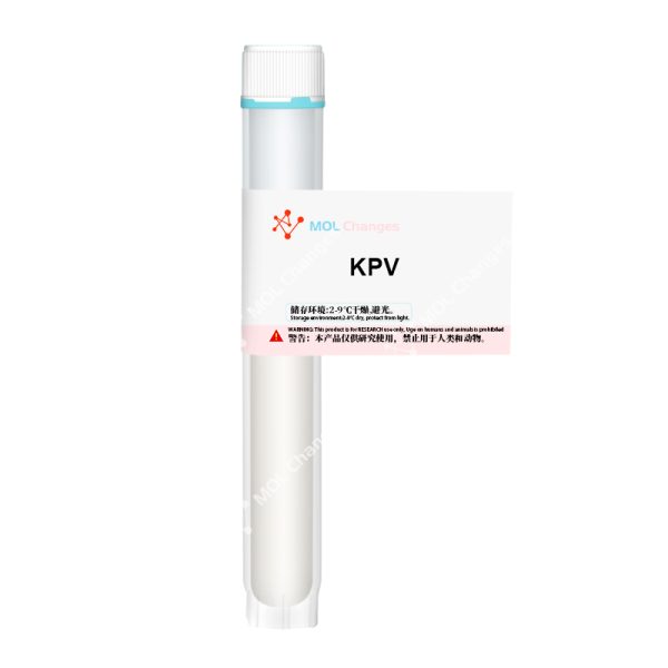
KPV Structure
- Sequence:Lys-Pro-Val
- CAS Number:67727-97-3
- Molecular Formula:C16H30N4O4
- Molecular Weight: 342.43 g/mol
What is KPV?
KPV is a C-terminal peptide fragment of α-melanocyte stimulating hormone (α-MSH). It is one of a number of short peptide derivatives of α-MSH that have been tested to determine whether they retain similar photoprotective properties, anti-ischemic activity, sexual effects, or benefits on feeding behavior and energy homeostasis.
KPV consists of lysine-proline-valine and has significant anti-inflammatory properties, and the peptide is actively being investigated as a potential therapy for inflammatory bowel disease. There is evidence of potent anti-inflammatory activity in the central nervous system, gastrointestinal tract, lungs, vascular system, and joints, and because KPV is a small peptide, it can be administered in a variety of ways, including orally, intravenously, and by transdermal routes.
KPV Effects
Gut Inflammation
Perhaps the most important finding in KPV research is that the peptide reduces intestinal inflammation. In a mouse model of inflammatory bowel disease (IBD), KPV showed robust results in reducing inflammatory infiltrates, MPO activity, and overall histologic evidence of inflammation, and mice treated with KPV in the study recovered faster and gained more weight than mice treated with placebo.
Further studies on the mechanism of KPV delivery have shown that loading KPV onto hyaluronic acid-functionalized nanoparticles helps to direct the inflammatory effects of the peptide to the appropriate sites within the gut. In mouse models, this accelerates mucosal healing and reduces inflammation by strongly down-regulating TNF-α. In many ways, KPV is a more effective and targeted approach to reduce IBD inflammation without affecting TNF-α elsewhere in the body. modifying KPV has the benefit of increasing the oral bioavailability of the peptide. This does not increase the efficacy of the peptide, but it does affect potency as well as the total dose required to achieve the effect.
KPV appears to be effective only in cases of excessive inflammation. It has little to no effect on normal tissue. This is due, at least in part, to the fact that KPV enters colon cells via transport proteins that are not regulated in inflammatory situations. This suggests that KPV may be an effective prophylactic or maintenance drug for IBD. It is safe to take even during the resting period because it has no effect. It is taken regularly so that the peptide is available when needed, otherwise it is excreted.Prof. Didier Merlin, who has led a number of studies on the potential gastrointestinal benefits of KPV, recently discovered that the peptide enters the colon cells via PepT1, a protein channel that is only expressed in any real quantity in the gut during inflammatory states. This helps explain why KPV is more effective in an already inflamed environment. It also suggests a new mode of drug delivery that can be applied to a wide range of conditions. By targeting proteins that are altered in disease conditions, even if they are not directly disease-causing, it is possible to focus the activity of a drug on certain regions. This could reduce the dosage of drugs with serious side effects and develop drugs that, although not effective in their own right, have a powerful therapeutic effect in the right disease state.
KPV as a general anti-inflammatory
As early as 1984, studies in rabbits showed KPV to be a powerful anti-inflammatory and antipyretic (fever reducer). However, in this case, KPV was less potent than the intact α-MSH molecule. This indicated to the scientists of the time that KPV lacked certain parts of the α-MSH molecule required for full antipyretic activity [5]. What followed were decades of research investigating various modified forms of α-MSH.
Perhaps the biggest lesson learned from these tests is that both α-MSH and several of its analogs reduce inflammation in a wide range of diseases. To date, these molecules have been tested in fever, irritant and allergic contact dermatitis, vasculitis, fibrosis, arthritis, and inflammation of the eyes, brain, lungs, and gastrointestinal tract. In all cases, alpha-MSH is the most effective anti-inflammatory agent. Unfortunately, it has one major side effect – it causes skin pigmentation. KPV, on the other hand, does not have this side effect. Although KPV is not as effective as intact α-MSH, its lack of side effects means that it is theoretically possible to increase levels to achieve the desired target effect in most cases.
Differences in potency have been found to be minimal at best, as most of the anti-inflammatory effects of α-MSH are actually due to the KPV fraction. Interestingly, however, the parent molecule seems to be more able to inhibit late inflammatory responses. For example, in contact dermatitis, α-MSH better prevents allergic inflammatory reactions up to 2 weeks after initial exposure. This suggests that α-MSH may influence some aspect of immune regulation that is not related to the immediate inflammatory response [7]. It is still being determined what this process is.
This figure shows ear swelling due to contact dermatitis at 24 hours (left) and 2 weeks (right). Note that at 24 hours, co-administration of KPV with the stimulus is almost as effective as co-administration of α-MSH with the stimulus. However, at 2 weeks, exposure to the irritant without co-administration of the peptide resulted in much less swelling of α-MSH compared to KPV.
Wound Healing
Wound healing is a complex physiological process. Scientists have identified three general stages of the wound healing process: inflammation, proliferation, and remodeling. Each phase is characterized by differences in cell populations and cytokine concentrations, representing a unique chemical/physiological environment for potential intervention. Studies have shown that although each stage of the wound healing process is characterized by different skin cell subtypes, the majority of cells express the melanocortin 1 receptor (MC1R), which binds α-melanocyte-stimulating hormone. Of course, this also means that these cell types also bind α-MSH analogs such as KPV and KdPT.
Since these α-MSH derivatives retain some of the properties of α-MSH but lack others, they are potentially beneficial in wound healing. For example, KPV provides the inflammatory properties of α-MSH but lacks the pigment-inducing activity of its parent peptide. This makes KPV a good candidate for improving wound healing while avoiding the characteristic skin changes normally associated with natural scar formation (a phenomenon that disproportionately affects darker-skinned individuals).
One of the reasons for the anti-inflammatory effects of KPV is its involvement in the innate immune response against two common skin pathogens. Studies have shown that KPV inhibits the growth of Staphylococcus aureus and Candida albicans. These benefits occur at physiologic concentrations, which means that KPV may provide an effective way to prevent infections in serious wounds such as burns.This benefit of KPV contrasts with other anti-inflammatory medications, which actually inhibit the body’s ability to fight infection. Thus, KPV combines anti-inflammatory activity with antimicrobial activity.
KPV is actually a structural model in recent studies aimed at replicating the antifungal effects of peptides in novel therapeutics. The idea is that the 3D structure of KPV makes it a potent antifungal agent, and replicating this structure could allow researchers to develop compounds that have the same antifungal activity but different effects on other biological processes.
Scar formation
Based on the known benefits of KPV in the first stage of wound healing (inflammation), the study also investigated its role in the other two stages of wound healing.KPV appears to reduce the chronic inflammation that leads to the formation of hypertrophic scars (e.g., scarring). This type of scar formation is characterized by extensive macrophage infiltration, TNF immunoreactivity and neutrophil abundance. Use of α-MSH in this setting results in smaller scars and a less dramatic inflammatory response. Similar effects have been found in other tissues such as the lungs and heart. These findings give hope that KPV can be used to prevent the scarring seen with certain chemotherapeutic drugs, which not only reduces the side effects of cancer treatments, but also increases the concentration of these drugs, thereby improving the efficacy of cancer treatments.
Referenced Citations
- M. E. Hiltz and J. M. Lipton, “Antiinflammatory activity of a COOH-terminal fragment of the neuropeptide alpha-MSH,” FASEB J. Off. Publ. Fed. Am. Soc. Exp. Biol., vol. 3, no. 11, pp. 2282–2284, Sep. 1989.
- K. Kannengiesser et al., “Melanocortin-derived tripeptide KPV has anti-inflammatory potential in murine models of inflammatory bowel disease,” Inflamm. Bowel Dis., vol. 14, no. 3, pp. 324–331, Mar. 2008, doi: 10.1002/ibd.20334.
- B. Xiao et al., “Orally Targeted Delivery of Tripeptide KPV via Hyaluronic Acid-Functionalized Nanoparticles Efficiently Alleviates Ulcerative Colitis,” Mol. Ther. J. Am. Soc. Gene Ther., vol. 25, no. 7, pp. 1628–1640, 05 2017, doi: 10.1016/j.ymthe.2016.11.020.
- G. Dalmasso, L. Charrier-Hisamuddin, H. T. T. Nguyen, Y. Yan, S. Sitaraman, and D. Merlin, “PepT1-Mediated Tripeptide KPV Uptake Reduces Intestinal Inflammation,” Gastroenterology, vol. 134, no. 1, pp. 166–178, Jan. 2008, doi: 10.1053/j.gastro.2007.10.026.
- D. B. Richards and J. M. Lipton, “Effect of alpha-MSH 11-13 (lysine-proline-valine) on fever in the rabbit,” Peptides, vol. 5, no. 4, pp. 815–817, Aug. 1984, doi: 10.1016/0196-9781(84)90027-5.
- T. Brzoska, T. A. Luger, C. Maaser, C. Abels, and M. Böhm, “Alpha-melanocyte-stimulating hormone and related tripeptides: biochemistry, antiinflammatory and protective effects in vitro and in vivo, and future perspectives for the treatment of immune-mediated inflammatory diseases,” Endocr. Rev., vol. 29, no. 5, pp. 581–602, Aug. 2008, doi: 10.1210/er.2007-0027.
- T. A. Luger and T. Brzoska, “α‐MSH related peptides: a new class of anti‐inflammatory and immunomodulating drugs,” Ann. Rheum. Dis., vol. 66, no. Suppl 3, pp. iii52–iii55, Nov. 2007, doi: 10.1136/ard.2007.079780.
- M. Cutuli, S. Cristiani, J. M. Lipton, and A. Catania, “Antimicrobial effects of alpha-MSH peptides,” J. Leukoc. Biol., vol. 67, no. 2, pp. 233–239, Feb. 2000, doi: 10.1002/jlb.67.2.233.
- M. F. Masman et al., “Synthesis and conformational analysis of His-Phe-Arg-Trp-NH2 and analogues with antifungal properties,” Bioorg. Med. Chem., vol. 14, no. 22, pp. 7604–7614, Nov. 2006, doi: 10.1016/j.bmc.2006.07.007.
- K. S. de Souza et al., “Improved cutaneous wound healing after intraperitoneal injection of alpha-melanocyte-stimulating hormone,” Exp. Dermatol., vol. 24, no. 3, pp. 198–203, Mar. 2015, doi: 10.1111/exd.12609.
- C. Lonati et al., “Modulatory effects of NDP-MSH in the regenerating liver after partial hepatectomy in rats,” Peptides, vol. 50, pp. 145–152, Dec. 2013, doi: 10.1016/j.peptides.2013.10.014.
- G. Colombo et al., “Gene expression profiling reveals multiple protective influences of the peptide alpha-melanocyte-stimulating hormone in experimental heart transplantation,” J. Immunol. Baltim. Md 1950, vol. 175, no. 5, pp. 3391–3401, Sep. 2005, doi: 10.4049/jimmunol.175.5.3391.
- G. Colombo et al., “Production and effects of alpha-melanocyte-stimulating hormone during acute lung injury,” Shock Augusta Ga, vol. 27, no. 3, pp. 326–333, Mar. 2007, doi: 10.1097/01.shk.0000239764.80033.7e.
- M. Schiller et al., “Human Dermal Fibroblasts Express Prohormone Convertases 1 and 2 and Produce Proopiomelanocortin-Derived Peptides,” J. Invest. Dermatol., vol. 117, no. 2, pp. 227–235, Aug. 2001, doi: 10.1046/j.0022-202x.2001.01412.x.
- T. Brzoska, M. Böhm, A. Lügering, K. Loser, and T. A. Luger, “Terminal signal: anti-inflammatory effects of α-melanocyte-stimulating hormone related peptides beyond the pharmacophore,” Adv. Exp. Med. Biol., vol. 681, pp. 107–116, 2010, doi: 10.1007/978-1-4419-6354-3_8.
- S. J. Getting, H. B. Schiöth, and M. Perretti, “Dissection of the anti-inflammatory effect of the core and C-terminal (KPV) alpha-melanocyte-stimulating hormone peptides,” J. Pharmacol. Exp. Ther., vol. 306, no. 2, pp. 631–637, Aug. 2003, doi: 10.1124/jpet.103.051623.
- K. Pawar, C. S. Kolli, V. K. Rangari, and R. J. Babu, “Transdermal Iontophoretic Delivery of Lysine-Proline-Valine (KPV) Peptide Across Microporated Human Skin,” J. Pharm. Sci., vol. 106, no. 7, pp. 1814–1820, Jul. 2017, doi: 10.1016/j.xphs.2017.03.017.
ALL ARTICLES AND PRODUCT INFORMATION PROVIDED ON THIS WEBSITE ARE FOR INFORMATONAL AND EDUCATIONAL PURPOSES ONLY.
In no way does this doctor/scientist endorse or advocate the purchase, sale, or use of this product for any reason. MOL Changes has no affiliation or relationship, implied or otherwise, with this physician. The purpose of citing this doctor is to acknowledge, acknowledge and commend the exhaustive research and development work done by the scientists working on this peptide.
HPLC test report
MS test report
Manufacturer Information
- KPV is manufactured by MOL Changes factory.
- KPV supplier MOL Changes.
- Maximum acceptable production volume: 100000 bottles.
- Content standard: net peptide.
- Purity: ≥98% for all products.
- Customization: 1mg-1g size customization is acceptable