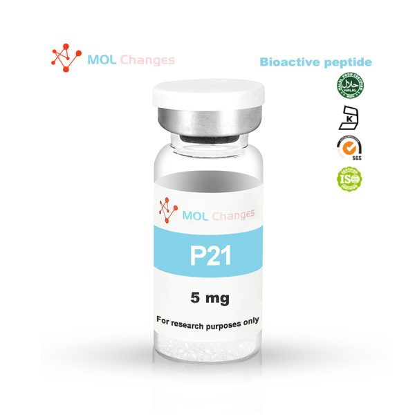
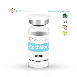
P21 is a modified, synthetic mimetic of CNTF. CNTF is a naturally occurring protein mediator of neuron growth in humans. The effects of CNTF have primarily been studied in the nervous system, though there are receptors for the peptide in other locations throughout the body (e.g. bone). It has been shown to promote neurotransmitter synthesis and neurite outgrowth. It also protects neurons and their supporting cells against inflammatory attacks. In addition to its neurotrophic effects, CNTF is known to increase satiety and thus reduce food intake.
CNTF and cerebrolysin are not the same molecule. P21 and cerebrolysin are not the same either. It is discussed below and contrasted with P21.
A recombinant version of CNTF was developed under the brand name Axokine. It was tested as a treatment for amyotrophic lateral sclerosis and is currently not sold. Interestingly, the body is quick to generate antibodies against Axokine, suggesting that there may be a potential for administering P21 and exogenous CNTF together in some settings, thus boosting CNTF levels while keeping antibody activity to a minimum.
Sequence
DGGL-adamantane-G
CAS Number
128-37-0
Molecular Formula
C30H54N6O5
Molecular Weight
987.55
P21 Related research
P21 is a small peptide derivative of CNTF. Small molecule mimics can perform some or all of the actions of larger neutral nutrient molecules without the side effects described above.P21 was developed through a process called epitope mapping, which uses antibodies to recognize the target binding site. For P21, antibodies directed against the CNTF receptor active site are first used to recognize the CNTF binding site. They were then used to identify which small synthetic peptides mimic CNTF binding and thus interfere with antibody binding . The result is P21, which not only binds to the CNTF receptor, but also crosses the blood-brain and placental/lactation barriers.P21 is a tetrapeptide derived from the most active region of the CNTF (amino acid residues 148-151).The C-terminal end of P21 has been added with an adamantylated glycine to increase the permeability of the blood-brain barrier and to reduce degradation by exopeptidases.
Natural CNTF is too large to cross the blood-brain barrier, has poor plasma stability, an unfavorable pharmacological profile, and actually promotes the production of anti-CNTF antibodies when administered systemically. Direct injection into the cerebrospinal fluid, while an option, is usually avoided due to pain, risk of infection, and other adverse effects. Unlike intact CNTF, P21 is stable in artificial gastric fluid for more than 30 minutes, which is sufficient to allow it to pass through the stomach in most cases. It is approximately 100% stable in the intestines for two hours, which is long enough for it to be absorbed. It is stable in plasma for more than 3 hours.
How does P21 work?
P21 has multiple roles in the CNS, but its primary role is in the dentate gyrus, where it acts to enhance neurogenesis and neuronal maturation in the granule cell layer and subgranular zone. The dentate gyrus is part of the hippocampal structure in the temporal lobe of the brain and is thought to contribute to the formation of new situational memories as well as spontaneous exploration/learning that occurs in new environments. The dentate gyrus also plays an important role in information preprocessing and pattern separation. Essentially, pattern separation allows mammals to distinguish one memory from another. The dentate gyrus is also of great interest to neuroscientists because it is one of the few brain regions known to have significant neurogenesis in adults.
Mouse modeling studies have shown that P21 does not bind to CNTF receptors, suggesting that despite its claim to be an analog, it should be clear that P21 is not a CNTF analog. It appears that P21 acts to inhibit antibodies or other molecules that neutralize CNTF. Thus, while P21 does not directly mimic the action of P21, it increases the concentration of this most potent neurogenesis enhancer, thereby effectively mimicking its action.
Studies in mice have shown that P21 increases the level of BrdU-positive cells in the dentate gyrus, a synthetic nucleoside (thymidine analog) used to detect proliferating cells in living tissue. In this experiment, it was found to be concentrated in the dentate gyrus of mice administered P21, but not in the DG of control mice, suggesting that P21 promotes cell proliferation in this region. To determine whether the cells are neurons, NeuN expression can be measured, as it is a marker for mature neurons. It is also significantly increased in mice administered P21 and in regions where BrdU is increased, supporting the idea that increased proliferation is actually increased neurogenesis.
Another component of P21 activity appears to be produced by inhibiting LIF-STAT signaling, an acronym for Leukemia Inhibitory Factor, a cytokine similar to interleukin 6 that plays an important role in embryogenesis. It is responsible for inhibiting differentiation and thus terminating cell proliferation in a controlled manner, a process that is important for enhancing tissue maturation, even if it comes at the cost of reduced proliferation. By inhibiting LIF, P21 removes one of the barriers to neurogenesis, thereby allowing the brain to enter an embryonic state more favorable for neuronal growth.
In Alzheimer’s disease (AD), the brain’s natural response to injury (i.e., neuronal and synaptic loss) is to increase activity in the dentate gyrus. Unfortunately, many aging brains lack the capacity to support neurogenesis, so alternative efforts fail.P21 Enhanced dentate gyrus activity is sufficient to overcome this limitation and help shift the balance of neurotrophic factors toward neurogenesis. Thus, limiting amyloid deposition in the brain may not be the only way to address the effects of AD. This could explain why, although plaque deposition begins early in AD, the effects of deposition do not become apparent until later in life, when neurotrophic factor homeostasis is shifted away from neurogenesis. It has been shown that neurotrophic support with P21 increases levels of brain-derived neurotrophic factor and neurotrophic protein 4, while decreasing the mitogenic effects of fibroblast growth factor 2. Interestingly, in a mouse model of AD, administration of P21 prior to the onset of AD prevented the cognitive decline that normally occurs. This suggests that P21 may be more important as a preventive measure than as a potential treatment.
It is also important to note that BDNF has been linked not only to enhanced neurogenesis, but also to the downregulation of certain enzymes responsible for tau protein and amyloid plaque formation in the AD brain. Specifically, BDNF decreases the activity of the GSK3-β protein, which catalyzes the formation of amyloid β from amyloid precursor proteins as well as the phosphorylation of tau proteins, which are steps in the development of AD that lead to inflammation and ultimately neurodegeneration.
It is worth pointing out that overproduction of GSK-3beta has been implicated in a number of disease processes, including type 2 diabetes, several different forms of cancer, and bipolar disorder. p21 and other GSK-3beta inhibitors are expected to play a role in the treatment of stroke, cancer, and especially bipolar disorder.
In particular, P21 appears to rescue the trend of decreased expression of MAP2 (microtubule-associated protein 2), a marker of synaptic growth between neurons, in AD-affected brains. Reduced levels of this protein are indicative of reduced synaptogenesis/neurogenesis and are a marker of disease progression in AD. Similarly, P21 is thought to rescue the decline in
- – Synaptic protein I, a key protein in synaptic communication between neurons.
- – GluR1 (AMPA receptor), a receptor that mediates rapid synaptic transmission.
- – NR1, a glutamate receptor associated with synaptic plasticity and learning.
Perhaps the most interesting thing about P21’s effect on synaptic proteins I, GluR1, and NR1 is that it can elevate these substances to supraphysiologic levels in both diseased and healthy brains. This led the researchers to conclude that P21 may not only help restore function in diseased brains, but also help enhance function in normal brains. As such, it may be useful as an intellectual stimulant and performance enhancer for cognitive tasks. Research in this area has not yet been conducted in animal models, let alone human trials. In fact, P21 is so effective in promoting neurogenesis that it actually increases the level of neurogenesis in diseased brains compared to healthy, untreated brains.
COA
HPLC
MS
- “Epitope Mapping – an overview | ScienceDirect Topics.” https://www.sciencedirect.com/topics/neuroscience/epitope-mapping (accessed Aug. 24, 2020).
- N. Baazaoui and K. Iqbal, “Prevention of dendritic and synaptic deficits and cognitive impairment with a neurotrophic compound,” Alzheimers Res. Ther., vol. 9, Jun. 2017, doi: 10.1186/s13195-017-0273-7.
- B. Li et al., “Neurotrophic peptides incorporating adamantane improve learning and memory, promote neurogenesis and synaptic plasticity in mice,” FEBS Lett., vol. 584, no. 15, pp. 3359–3365, 2010, doi: 10.1016/j.febslet.2010.06.025.
- S. F. Kazim et al., “Disease modifying effect of chronic oral treatment with a neurotrophic peptidergic compound in a triple transgenic mouse model of Alzheimer’s disease,” Neurobiol. Dis., vol. 71, pp. 110–130, Nov. 2014, doi: 10.1016/j.nbd.2014.07.001.
- S. F. Kazim and K. Iqbal, “Neurotrophic factor small-molecule mimetics mediated neuroregeneration and synaptic repair: emerging therapeutic modality for Alzheimer’s disease,” Mol. Neurodegener., vol. 11, Jul. 2016, doi: 10.1186/s13024-016-0119-y.
- J. J. Luykx, M. P. M. Boks, A. P. R. Terwindt, S. Bakker, R. S. Kahn, and R. A. Ophoff, “The involvement of GSK3β in bipolar disorder: Integrating evidence from multiple types of genetic studies,” Eur. Neuropsychopharmacol., vol. 20, no. 6, pp. 357–368, Jun. 2010, doi: 10.1016/j.euroneuro.2010.02.008.
- M. K. Mohammad et al., “Olanzapine inhibits glycogen synthase kinase-3beta: an investigation by docking simulation and experimental validation,” Eur. J. Pharmacol., vol. 584, no. 1, pp. 185–191, Apr. 2008, doi: 10.1016/j.ejphar.2008.01.019.
- B. Xu and X. Xie, “Neurotrophic factor control of satiety and body weight,” Nat. Rev. Neurosci., vol. 17, no. 5, pp. 282–292, May 2016, doi: 10.1038/nrn.2016.24.
- K. Iqbal, S. F. Kazim, S. Bolognin, and J. Blanchard, “Shifting balance from neurodegeneration to regeneration of the brain: a novel therapeutic approach to Alzheimer’s disease and related neurodegenerative conditions,” Neural Regen. Res., vol. 9, no. 16, pp. 1518–1519, Aug. 2014, doi: 10.4103/1673-5374.139477.
- “Rat and Mice Weights | Animal Resources Centre.” http://www.arc.wa.gov.au/?page_id=125 (accessed Aug. 26, 2020).
- E. Rockenstein, M. Mante, A. Adame, L. Crews, H. Moessler, and E. Masliah, “Effects of Cerebrolysin on neurogenesis in an APP transgenic model of Alzheimer’s disease,” Acta Neuropathol. (Berl.), vol. 113, no. 3, pp. 265–275, Mar. 2007, doi: 10.1007/s00401-006-0166-5.
- H. Chen, Y.-C. Tung, B. Li, K. Iqbal, and I. Grundke-Iqbal, “Trophic factors counteract elevated FGF-2-induced inhibition of adult neurogenesis,” Neurobiol. Aging, vol. 28, no. 8, pp. 1148–1162, Aug. 2007, doi: 10.1016/j.neurobiolaging.2006.05.036.
Article/Literature Citation Notes
The purpose of quoting the scientist and professor's article is to acknowledge, recognise and applaud the exhaustive development work that has been done to undertake this peptide research. The scientist does not in any way support or advocate the purchase, sale or use of this product for any reason, and MOL Changes has no affiliation or relationship, implied or otherwise, with the scientist.
warning
These products are intended for scientific purposes only for in vitro research and are not to be used for human, animal or unethical experiments, in vitro research (Latin: in glass) is conducted in vitro. These products are not drugs and are not approved by the Food and Drug Administration in any country for the prevention, treatment or cure of any medical condition, disease or illness. The introduction of this product into humans or animals in any form is strictly prohibited by law.


