
Tali vave
Fo'ou 2026 pore-formation research shows why purity certificates can't predict membrane activity, and which functional assays close the gap.
Why the Conventional Purity-First View of Pore-Forming Peptide Design Is Incomplete

The mainstream release criterion for a membrane-active peptide is a single number: reverse-phase HPLC area purity, usually at or above 95%, plus a mass spectrum confirming the expected intact mass. That standard is genuinely useful, and it is worth being precise about what it establishes. RP-HPLC reports chromatographic purity, meaning the share of UV-absorbing material sitting in the main peak, while mass spectrometry reports intact mass and, mo e MS/MS, sequence-level fragmentation support (PMC methods chapter). Together they answer a real question: what was made, and how pure is it.
The tiers themselves are conventions rather than requirements. Suppliers commonly quote ≥95% RP-HPLC area as the research-grade floor, ≥98% as the modern research and GMP floor, and ≥99% for premium or clinical material (peptide purity reference set, 2025). These are reported supplier conventions, not a compendial mandate.

The view became dominant because it is cheap, fast, fakahangatonu, and sufficient for most non-membrane targets. Its origin is analytical-chemistry practice, not biophysics, which is where the first peptide purity testing limitations appear: percent-area purity does not name the impurities it excludes.
⚠️ Fakatokanga: percent-area purity does not quantify deletion sequences, ngaahi fakanounou, co-eluting diastereomers, ngaahi fakafepaki (TFA, ʻaseiti, kololaiti) or water content, so net peptide content can differ from label weight (peptide purity-testing guide, 2026).

Fakatahataha'i 'o e Peptide Two samples of equal nominal mass can therefore carry different effective concentrations, and neither certificate tells you whether the peptide inserts, oligomerizes, or forms a stable pore in a membrane.
Ko e hā ʻa e . 2026 Pore-Formation Research Actually Shows
That reframing starts with the 2026 Natula Paiolosia Kemikale study from Deb and colleagues, which treats pore-forming peptide design as a sequence-encoded property rather than a purification outcome. The team ran 51 µs of unbiased coarse-grained molecular dynamics (Martini 2.2 / GROMACS) kolosi fakafuofua 150 pre-assembled octameric transmembrane barrel pores in POPC, validated the trajectories with 2 µs all-atom simulations, then synthesized 22 peptides and tested them by calcein leakage, planar bilayer electrophysiology, real-time AFM, CD, and GUV dextran transport (Ko e Sosaieti Peptide ʻAmelika, 2026-08-06).
Two results matter for anyone editing a sequence. ʻuluaki, conductance arrived in discrete steps: single-pore values of 0.116, 0.251, 0.353, mo e 0.424 nS (Ko e Sosaieti Peptide ʻAmelika, 2026-08-06). A barrel-stave pore, where peptides line a fixed channel, and a toroidal pore, where the lipid bilayer itself curves into the lining, produce different conductance signatures, so the stepwise pattern is a readout of architecture, not just of how much peptide bound. Fika ua, real-time AFM caught pores roughly 6 nm wide within 20 minutes of peptide addition (Ko e Sosaieti Peptide ʻAmelika, 2026-08-06).
The pipeline also distilled 52 permissible sequence templates in four classes, ranked by interaction density from a 150-peptide MD scan (Ko e Sosaieti Peptide ʻAmelika, 2026-08-06). Read those numbers as assay conditions, not constants: each conductance value belongs to a specific bilayer model, malohi ionic, and voltage, and none of them transfers directly to a different membrane system.
How Sequence Edits Move a Peptide Between Pore Phenotypes

The unit that changes peptide sequence and membrane activity is the substitution, 'ikai ko e peptide .. Single and clustered edits can move one backbone between three states: no membrane activity, transient leakage, and a stable pore.
Aromatic placement is the clearest lever. In Deb et al.’s designs, optimal aromatic positions shifted from residues 13 mo e 19 in a 30-residue core to 13, 15, 8 mo e 26 when the core was shortened to 22 toenga, and helix tilt changed from 50° to 30° relative to the membrane normal (Ko e Sosaieti Peptide ʻAmelika, 2026-08-06). Charge can substitute for that anchoring: four Arg–Asp salt bridges per interface retained pore integrity even with aromatic stacking removed (Ko e Sosaieti Peptide ʻAmelika, 2026-08-06). Electrostatics set the driving force, since cationic peptides showed roughly 20 kJ mol⁻¹ greater affinity for anionic POPE:POPG than zwitterionic POPC bilayers (Ko e Sosaieti Peptide ʻAmelika, 2026-08-06).
Stereochemistry is an independent second axis. Swapping every residue of DpPorA to its D-enantiomer changed unitary conductance from about 4 nS to 1.5 ± 0.3 nS at +100 mV in 1 M KCl (n = 75), and across 100 insertions the D-form split 75%/25% between the ~1.5 nS and ~4 nS states with PK⁺/PCl⁻ ≈ 7:1 (Ge et al., Nat Angamaheni, 2022-09-14).
|
Fotunga hokohoko |
Expected membrane effect |
Assay that detects it |
|---|---|---|
|
Helicity |
Whether the peptide can span the bilayer as a continuous helix |
Circular dichroism in POPC vs POPE:POPG vesicles |
|
Hydrophobic moment |
Depth and stability of membrane insertion |
Tryptophan fluorescence shift; vesicle leakage kinetics |
|
Charge placement |
Lipid-headgroup selectivity and salt-bridge stabilization |
Zeta potential; conductance in anionic vs zwitterionic bilayers |
|
Aromatic placement |
Interfacial anchoring and helix tilt |
Oriented circular dichroism; solid-state NMR tilt angles |
|
Geometry and tilt |
Which architecture forms: barrel-stave, toroidal or β-sheet |
Single-channel conductance; oriented CD; FTIR amide I |
Lisini, arginine and bulkier residues act as membrane-desorption gatekeepers, blocking deep insertion or bulk translocation (PMC, 2021-12-24).
Why Purity Testing Cannot Predict Membrane Behavior
You have probably seen this already: two vials, same nominal mass, same certificate, and one of them simply does not behave the way the assay says it should. The certificate is not wrong. It is answering a narrower question than the one you are asking.
Reverse-phase HPLC and mass spectrometry establish identity and chromatographic purity. Neither establishes fold or secondary structure, tu'unga 'olikomeli, aggregation state under native conditions, or membrane behaviour; those require separate biophysical or cell-based assays (PMC analytical review). Percent-area purity also carries a counterion and residual-water confound, so two samples of equal nominal mass can deliver different effective concentrations once dissolved. Different effective concentration means different apparent membrane activity, even when the sequence is identical.
Functional results are noisy for their own reasons. Minimum inhibitory concentration values commonly move by one to two dilution steps between laboratories, users and methods, and omitting controls, using the wrong inoculum (specified as 5 × 10⁵ CFU/mL) or deviating from growth conditions can distort results (MIC methods review, 2024). The reproducibility problem extends to prediction: ʻi ha 2026 benchmark of 48 published antimicrobial-peptide prediction methods, fewer than 25% were reproducible (BATTLE-AMP benchmark, bioRxiv, 2026). That is a preprint benchmark of methods, not of peptides, but it sets expectations for how much of a design decision can be settled on paper.
This is where custom peptide characterization ends and your own work begins.
Tefito'i 'o e 'ave: analytical release testing is vendor-side; membrane-function testing is customer-side.
A Better Framework for Pore-Forming Peptide Design
Design pore-forming peptides against a target pore architecture and a target assay, not against a purity specification. That single change in the design target reorganizes the whole project.
Five principles follow from the 2026 teita:
-
Choose the architecture first. Decide whether you want a barrel-stave, toroidal, or disordered-thinning pore before you touch the sequence, because each architecture implies a different set of sequence constraints.
-
Treat aromatic placement and salt bridges as the stability layer. They govern how long the assembly holds together in a membrane, not how strongly it binds.
-
Treat charge placement as the selectivity layer. Where the cationic residues sit relative to the hydrophobic face sets which membranes the peptide discriminates between.
-
Treat stereochemistry as a phenotype variable. A D-amino acid substitution is not a footnote; it can move a peptide between pore phenotypes.
-
Pre-commit to the functional readout before synthesis. Decide the assay, the lipid system, and the peptide-to-lipid ratios in advance, so the synthesis batch has something to be tested against.
The proof point is that this is workable at scale. HA 2026 design pipeline moved from a 150-peptide in-silico scan to 22 synthesized peptides, with calcein leakage and computed pore hydration correlating across the set. The lead peptide, KDFA2i + 9-NH2, produced roughly 20% calcein release at a peptide-to-lipid ratio of 1:2,000, rising at higher ratios (Ko e Sosaieti Peptide ʻAmelika, 2026). Note what that number is: a functional result at a defined ratio in a defined system, which is exactly the kind of evidence a certificate of analysis cannot supply.
For teams without in-house synthesis, MOL Changes supports custom sequence design and modification work, which can be used to turn a chosen architecture into an orderable peptide while the functional testing stays in your own assay.
None of this is deterministic. Sequence-to-pore rules are probabilistic, and some well-designed peptides will still fail their first assay. The framework’s value is that it makes the failure informative rather than ambiguous.
Choosing Functional Assays That Complement Purity Data
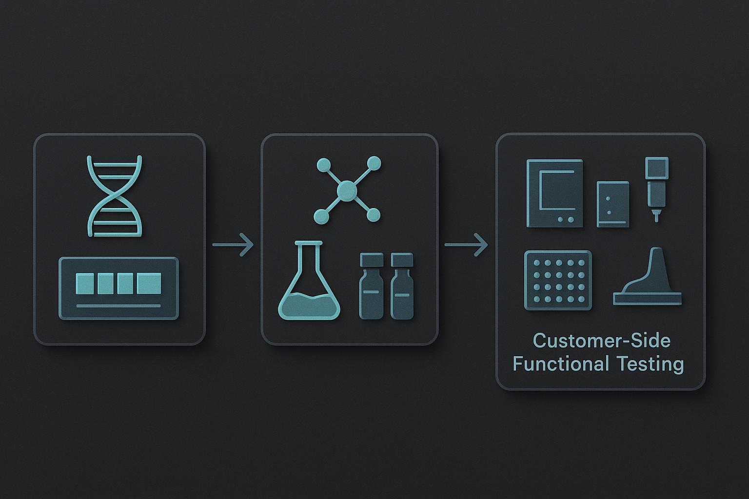
The first action step is to pick the assay that answers the question purity testing cannot: not “what is in this vial,” but “what does this peptide do to a membrane.” For Peptides fakatupu functional assays for membrane-active peptides, that means matching the readout to the pore property under test rather than defaulting to whichever plate reader is free.
Calcein leakage measures dye release from lipid vesicles and reports bulk permeabilization. DiSC₃-5 depolarization tracks the loss of a membrane potential across a bacterial membrane, so it reports membrane disruption in a more physiological context. Planar bilayer single-channel recording measures current through one pore at a time, which is the only one of the three that resolves unitary conductance.
The three sit on very different sensitivity scales. Calcein leakage typically needs peptide concentrations in the 1 ki 25 µM range against roughly 20 µM lipid with 75 mM intraliposomal calcein, while DiSC₃-5 depolarization effects cluster around 19 μM. Single-channel work reaches sub-micromolar to low-micromolar concentrations, with published detections at 0.3 μM, 0.8 ki 4.1 μM, mo e 6.2 μM, resolving conductances from about 6 ki 360 pS up to the nanosiemens range. A peptide that looks inert in a leakage assay may simply be below the detection floor of that readout.
|
Assay |
Fehu'i 'oku ne tali . |
Required control |
Blind spot |
|---|---|---|---|
|
Calcein leakage |
Does the peptide permeabilize lipid bilayers in bulk? |
Vesicles without peptide; detergent for 100% 'atā |
Averages away transient or rare pores |
|
DiSC₃-5 depolarization |
Does it collapse membrane potential? |
Protonophore control; peptide-free membrane |
Cannot separate pore formation from membrane solubilization |
|
Planar bilayer single-channel |
What is the unitary conductance and lifetime of one pore? |
Peptide-free bilayer; voltage-reversal runs |
Low throughput; one pore may not represent the population |
The blind spot in the first row is not hypothetical. A Wza-based D-peptide, DcWza, produced unitary conductance of 0.95 ± 0.1 nS at +200 mV in 1 M KCl across 50 ngaahi meʻa naʻe hokó, yet vesicle permeabilization after one hour was 4.6 ± 2.5% against a control of 4.2 ± 2.1% (Ge et al., Fetu'utaki 'a Natula, 2022). Transient pores that eject rather than persist leave almost no trace in a bulk leakage readout. That is the case for running single-channel work alongside, not instead of, vesicle assays.
Implementation, ʻi he hokohoko:
-
Define the membrane model and lipid composition. Match the lipid mix to the biological membrane you care about. Ikuna vave: a day of reading and a decision.
-
Select the readout. Bulk leakage for a first pass, depolarization for a physiological context, single-channel for mechanism. Taimi loloa ange: single-channel rigs take weeks to commission.
-
Fix the controls. Peptide-free vesicles, a positive disruptor, and a solvent-only blank. Ikuna vave, but skipping it invalidates everything downstream.
-
Set the concentration range. Span at least two orders of magnitude around your expected threshold, and remember the detection floors above.
-
Pre-register the pass criterion. Write down what counts as active before you run the plate. This is the step most teams skip and most regret.
Measure leakage rate, unitary conductance, and selectivity ratio. A realistic timeline is two to four weeks for a validated vesicle assay and two to three months before single-channel data is publication-grade.
Caveats and Where the Conventional View Still Holds
The design rules are probabilistic, not deterministic, and some designed pores fail. That is the most important limitation to state plainly, because it is also the honest reason purity-first thinking persists: purity is measurable, repeatable, and cheap to certify, while membrane behavior is none of those things. Ngaohi 'o e Peptide
Context matters more than the argument does. For non-membrane targets, or for screening libraries where the readout is binding rather than permeabilization, RP-HPLC and MS purity genuinely is the right release criterion, and peptide purity testing limitations only become a problem when that certificate is asked to answer a question it was never designed to answer.
The weakest part of the case is the evidence base itself. Much of the 2026 design guidance derives from a single coarse-grained MD pipeline run in POPC, and the salt-bridge rescue was demonstrated in simulation before anyone synthesized the peptide. Neither result is wrong; both are narrower than a design rule implies.
Treat the framework as a hypothesis generator, 'ikai ko ha fakaikiiki. It tells you which sequence edits are worth making and which assays are worth running, and it still expects some of those pores to fail.
But Doesn’t High Purity Guarantee Reproducible Membrane Activity?
ʻIkai. Purity constrains what is in the vial, not how that material assembles in a bilayer. A ≥98% specification is not meaningless, ka: it removes a large class of confounds, including truncated sequences, tamate'i 'a e ngaahi me'a 'uli, and scavenger by-products that would otherwise muddy any activity readout. What it cannot do is tell you whether the peptide inserts, oligomerizes, or forms the pore architecture your design assumed.
If a project is already locked into a purity-only release workflow, you do not need to rebuild the program. Add one orthogonal functional readout, such as a vesicle leakage assay or a planar bilayer conductance measurement, and use it to confirm that the released lot behaves as the design intended.
Treat activity reported from a peptide characterized only by HPLC and MS as provisional. Without visible assay conditions and controls, you cannot tell whether the observed effect came from the peptide, a residual counterion, or the buffer.
Aofangatuku: From Purity Certificate to Functional Evidence
Purity is necessary but not sufficient for pore-forming peptide design: a chromatogram can confirm what a molecule is, never what it does at a membrane.
The practical shift is in release criteria. For membrane-active peptides, analytical data should travel with at least one functional readout, and published activity numbers should arrive alongside the assay conditions and controls that produced them. That pairing is what makes a result reproducible in someone else’s hands rather than only in the lab that generated it.
None of this asks you to rebuild an existing program. Start by mapping the sequence features you already control onto the membrane behavior you actually need, then pick the assay that detects that behavior. A design workflow and characterization checklist can make that mapping explicit before the next lot is ordered.
The direction of travel is toward design rules that are probabilistic but usable: good enough to narrow a sequence space, honest enough to say when a designed pore will not behave as intended.