
Por qué la visión convencional de pureza del diseño de péptidos formadores de poros está incompleta

El criterio de liberación principal para un péptido activo en la membrana es un número único.: pureza del área de HPLC de fase inversa, generalmente en o por encima 95%, más un espectro de masas que confirma la masa intacta esperada. Ese estándar es realmente útil., y vale la pena ser preciso sobre lo que establece. RP-HPLC informa la pureza cromatográfica, es decir, la proporción de material absorbente de rayos UV que se encuentra en el pico principal, mientras que la espectrometría de masas informa masa intacta y, con MS/MS, soporte de fragmentación a nivel de secuencia (Capítulo de métodos PMC). Juntos responden a una pregunta real: lo que se hizo, y que puro es.
Los niveles en sí son convenciones más que requisitos.. Los proveedores suelen citar ≥95 % de área de RP-HPLC como piso de grado de investigación, ≥98% como piso de investigación moderno y GMP, y ≥99% para material premium o clínico (conjunto de referencia de pureza de péptidos, 2025). Estas son convenciones de proveedores informadas., no es un mandato compendial.

La vista se volvió dominante porque es barata., rápido, ortogonal, y suficiente para la mayoría de objetivos que no son de membrana. Su origen es la práctica de la química analítica., no biofísica, Aquí es donde aparecen las primeras limitaciones en las pruebas de pureza de péptidos.: La pureza porcentual del área no nombra las impurezas que excluye..
⚠️ Advertencia: la pureza porcentual de área no cuantifica las secuencias de eliminación, truncamientos, diastereómeros coeluyentes, contraiones (TFA, acetato, cloruro) o contenido de agua, por lo que el contenido neto de péptidos puede diferir del peso de la etiqueta (guía de prueba de pureza de péptidos, 2026).

Síntesis de péptidos Por lo tanto, dos muestras de igual masa nominal pueden contener diferentes concentraciones efectivas., y ninguno de los certificados le indica si el péptido se inserta, oligomeriza, o forma un poro estable en una membrana.
¿Qué 2026 La investigación sobre la formación de poros realmente lo demuestra
Ese replanteamiento comienza con la 2026 Naturaleza Química Biología estudio de Deb y colegas, que trata el diseño de péptidos formadores de poros como una propiedad codificada por secuencia en lugar de un resultado de purificación. el equipo corrió 51 µs de dinámica molecular imparcial de grano grueso (martini 2.2 / GROMACS) aproximadamente 150 poros de barril transmembrana octamérica preensamblados en POPC, validó las trayectorias con 2 µs simulaciones de todos los átomos, luego sintetizado 22 péptidos y los probó mediante fuga de calceína., electrofisiología bicapa plana, AFM en tiempo real, CD, y transporte de dextrano GUV (Sociedad Americana de Péptidos, 2026-08-06).
Dos resultados son importantes para cualquiera que edite una secuencia. Primero, la conductancia llegó en pasos discretos: valores de poro único de 0.116, 0.251, 0.353, y 0.424 nS (Sociedad Americana de Péptidos, 2026-08-06). Un poro de duela de barril, donde los péptidos recubren un canal fijo, y un poro toroidal, donde la bicapa lipídica se curva hacia el revestimiento, producir diferentes firmas de conductancia, entonces el patrón paso a paso es una lectura de la arquitectura, no sólo de cuánto péptido se une. Segundo, AFM en tiempo real atrapó los poros con brusquedad 6 nm de ancho dentro 20 minutos de adición de péptidos (Sociedad Americana de Péptidos, 2026-08-06).
El oleoducto también destiló 52 plantillas de secuencia permitidas en cuatro clases, clasificados por densidad de interacción a partir de una exploración MD de 150 péptidos (Sociedad Americana de Péptidos, 2026-08-06). Lea esos números como condiciones de ensayo., no constantes: cada valor de conductancia pertenece a un modelo bicapa específico, fuerza iónica, y voltaje, y ninguno de ellos se transfiere directamente a un sistema de membrana diferente.
Cómo las ediciones de secuencia mueven un péptido entre fenotipos de poros

La unidad que cambia la secuencia peptídica y la actividad de la membrana es la sustitución., no el péptido. Las ediciones individuales y agrupadas pueden mover una columna vertebral entre tres estados: sin actividad de membrana, fuga transitoria, y un poro estable.
La colocación aromática es la palanca más clara.. En los diseños de Deb et al., posiciones aromáticas óptimas desplazadas de los residuos 13 y 19 en un núcleo de 30 residuos para 13, 15, 8 y 26 cuando el núcleo fue acortado a 22 residuos, y la inclinación de la hélice cambió de 50° a 30° en relación con la membrana normal (Sociedad Americana de Péptidos, 2026-08-06). La carga puede sustituir ese anclaje: cuatro puentes salinos Arg-Asp por interfaz mantuvieron la integridad de los poros incluso sin el apilamiento aromático (Sociedad Americana de Péptidos, 2026-08-06). La electrostática establece la fuerza motriz, ya que los péptidos catiónicos mostraron aproximadamente 20 kJ mol⁻¹ mayor afinidad por el POPE aniónico:POPG que las bicapas zwitteriónicas de POPC (Sociedad Americana de Péptidos, 2026-08-06).
La estereoquímica es un segundo eje independiente.. El intercambio de cada residuo de DpPorA por su enantiómero D cambió la conductancia unitaria de aproximadamente 4 nS a 1.5 ± 0.3 nS en +100 mV en 1 KCl (norte = 75), y a través 100 inserciones de la división en forma de D 75%/25% entre los estados ~1,5 nS y ~4 nS con PK⁺/PCl⁻ ≈ 7:1 (Ge et al., Nat común, 2022-09-14).
|
Característica de secuencia |
Expected membrane effect |
Assay that detects it |
|---|---|---|
|
Helicity |
Whether the peptide can span the bilayer as a continuous helix |
Circular dichroism in POPC vs POPE:POPG vesicles |
|
Momento hidrofóbico |
Depth and stability of membrane insertion |
Tryptophan fluorescence shift; vesicle leakage kinetics |
|
Charge placement |
Lipid-headgroup selectivity and salt-bridge stabilization |
Zeta potential; conductance in anionic vs zwitterionic bilayers |
|
Aromatic placement |
Interfacial anchoring and helix tilt |
Oriented circular dichroism; solid-state NMR tilt angles |
|
Geometry and tilt |
Which architecture forms: barrel-stave, toroidal or β-sheet |
Single-channel conductance; oriented CD; FTIR amide I |
lisina, arginine and bulkier residues act as membrane-desorption gatekeepers, blocking deep insertion or bulk translocation (PMC, 2021-12-24).
Por qué las pruebas de pureza no pueden predecir el comportamiento de la membrana
You have probably seen this already: two vials, same nominal mass, same certificate, and one of them simply does not behave the way the assay says it should. The certificate is not wrong. It is answering a narrower question than the one you are asking.
Reverse-phase HPLC and mass spectrometry establish identity and chromatographic purity. Neither establishes fold or secondary structure, estado oligomerico, aggregation state under native conditions, or membrane behaviour; those require separate biophysical or cell-based assays (PMC analytical review). Percent-area purity also carries a counterion and residual-water confound, so two samples of equal nominal mass can deliver different effective concentrations once dissolved. Different effective concentration means different apparent membrane activity, even when the sequence is identical.
Functional results are noisy for their own reasons. Minimum inhibitory concentration values commonly move by one to two dilution steps between laboratories, users and methods, and omitting controls, using the wrong inoculum (specified as 5 × 10⁵ CFU/mL) or deviating from growth conditions can distort results (MIC methods review, 2024). The reproducibility problem extends to prediction: en un 2026 benchmark of 48 published antimicrobial-peptide prediction methods, fewer than 25% were reproducible (BATTLE-AMP benchmark, bioRxiv, 2026). That is a preprint benchmark of methods, not of peptides, but it sets expectations for how much of a design decision can be settled on paper.
This is where custom peptide characterization ends and your own work begins.
Conclusión clave: analytical release testing is vendor-side; membrane-function testing is customer-side.
Un mejor marco para el diseño de péptidos formadores de poros
Design pore-forming peptides against a target pore architecture and a target assay, not against a purity specification. That single change in the design target reorganizes the whole project.
Five principles follow from the 2026 datos:
-
Choose the architecture first. Decide whether you want a barrel-stave, toroidal, or disordered-thinning pore before you touch the sequence, because each architecture implies a different set of sequence constraints.
-
Treat aromatic placement and salt bridges as the stability layer. They govern how long the assembly holds together in a membrane, not how strongly it binds.
-
Treat charge placement as the selectivity layer. Where the cationic residues sit relative to the hydrophobic face sets which membranes the peptide discriminates between.
-
Treat stereochemistry as a phenotype variable. A D-amino acid substitution is not a footnote; it can move a peptide between pore phenotypes.
-
Pre-commit to the functional readout before synthesis. Decide the assay, the lipid system, and the peptide-to-lipid ratios in advance, so the synthesis batch has something to be tested against.
The proof point is that this is workable at scale. A 2026 design pipeline moved from a 150-peptide in-silico scan to 22 synthesized peptides, with calcein leakage and computed pore hydration correlating across the set. The lead peptide, KDFA2i + 9-NH₂, produced roughly 20% calcein release at a peptide-to-lipid ratio of 1:2,000, rising at higher ratios (Sociedad Americana de Péptidos, 2026). Note what that number is: a functional result at a defined ratio in a defined system, which is exactly the kind of evidence a certificate of analysis cannot supply.
For teams without in-house synthesis, MOL Changes supports custom sequence design and modification work, which can be used to turn a chosen architecture into an orderable peptide while the functional testing stays in your own assay.
None of this is deterministic. Sequence-to-pore rules are probabilistic, and some well-designed peptides will still fail their first assay. The framework’s value is that it makes the failure informative rather than ambiguous.
Elección de ensayos funcionales que complementen los datos de pureza
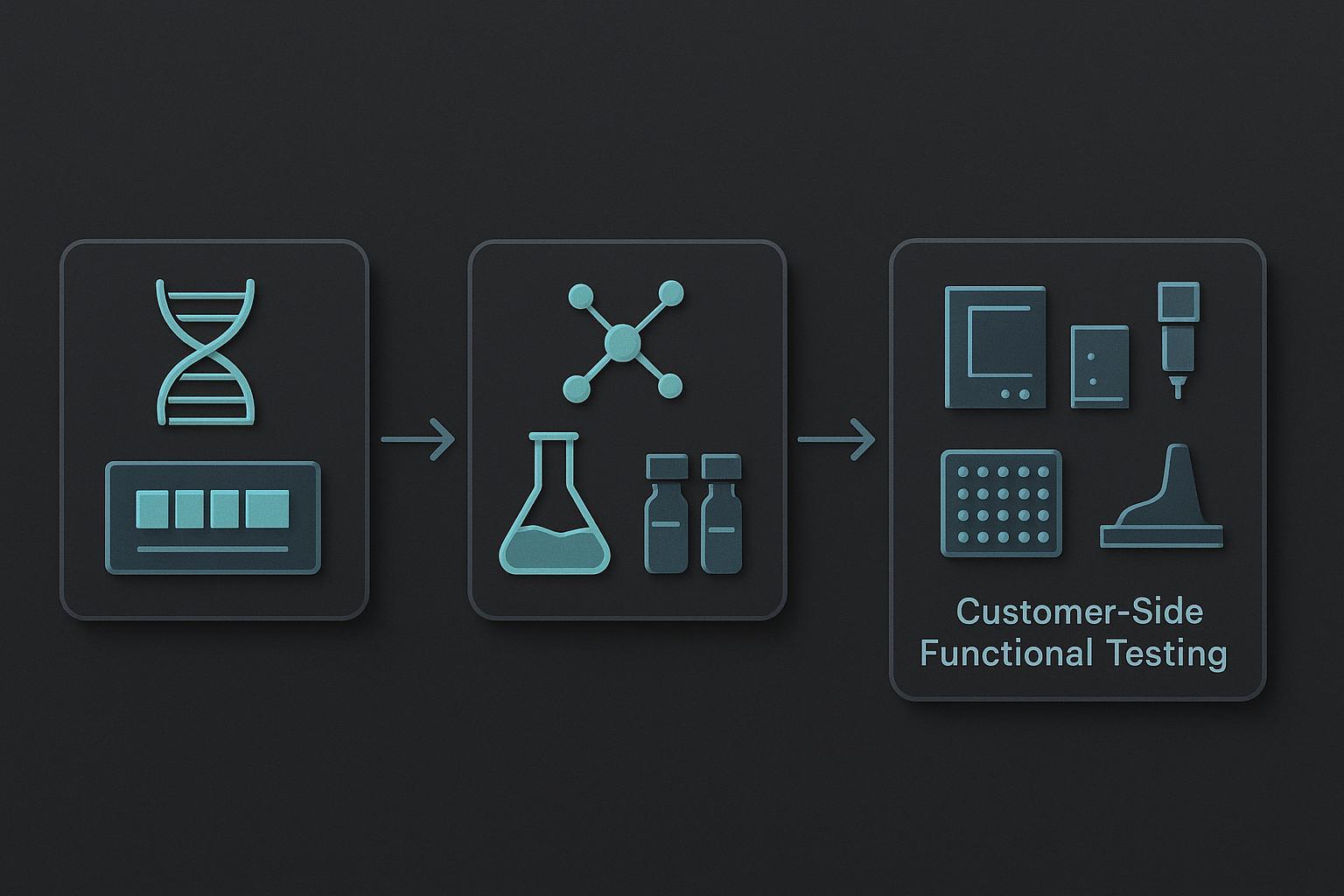
The first action step is to pick the assay that answers the question purity testing cannot: not “what is in this vial,” but “what does this peptide do to a membrane.” For Péptidos sintéticos functional assays for membrane-active peptides, that means matching the readout to the pore property under test rather than defaulting to whichever plate reader is free.
Calcein leakage measures dye release from lipid vesicles and reports bulk permeabilization. DiSC₃-5 depolarization tracks the loss of a membrane potential across a bacterial membrane, so it reports membrane disruption in a more physiological context. Planar bilayer single-channel recording measures current through one pore at a time, which is the only one of the three that resolves unitary conductance.
The three sit on very different sensitivity scales. Calcein leakage typically needs peptide concentrations in the 1 a 25 µM range against roughly 20 µM lipid with 75 mM intraliposomal calcein, while DiSC₃-5 depolarization effects cluster around 19 µM. Single-channel work reaches sub-micromolar to low-micromolar concentrations, with published detections at 0.3 µM, 0.8 a 4.1 µM, y 6.2 µM, resolving conductances from about 6 a 360 pS up to the nanosiemens range. A peptide that looks inert in a leakage assay may simply be below the detection floor of that readout.
|
Ensayo |
Question it answers |
Required control |
Blind spot |
|---|---|---|---|
|
Calcein leakage |
Does the peptide permeabilize lipid bilayers in bulk? |
Vesicles without peptide; detergent for 100% liberar |
Averages away transient or rare pores |
|
DiSC₃-5 depolarization |
Does it collapse membrane potential? |
Protonophore control; peptide-free membrane |
Cannot separate pore formation from membrane solubilization |
|
Planar bilayer single-channel |
What is the unitary conductance and lifetime of one pore? |
Peptide-free bilayer; voltage-reversal runs |
Low throughput; one pore may not represent the population |
The blind spot in the first row is not hypothetical. A Wza-based D-peptide, DcWza, produced unitary conductance of 0.95 ± 0.1 nS en +200 mV en 1 M KCl across 50 events, yet vesicle permeabilization after one hour was 4.6 ± 2.5% against a control of 4.2 ± 2.1% (Ge et al., Comunicaciones de la naturaleza, 2022). Transient pores that eject rather than persist leave almost no trace in a bulk leakage readout. That is the case for running single-channel work alongside, not instead of, vesicle assays.
Implementation, in order:
-
Define the membrane model and lipid composition. Match the lipid mix to the biological membrane you care about. Quick win: a day of reading and a decision.
-
Select the readout. Bulk leakage for a first pass, depolarization for a physiological context, single-channel for mechanism. Longer-term: single-channel rigs take weeks to commission.
-
Fix the controls. Peptide-free vesicles, a positive disruptor, and a solvent-only blank. Quick win, but skipping it invalidates everything downstream.
-
Set the concentration range. Span at least two orders of magnitude around your expected threshold, and remember the detection floors above.
-
Pre-register the pass criterion. Write down what counts as active before you run the plate. This is the step most teams skip and most regret.
Measure leakage rate, unitary conductance, and selectivity ratio. A realistic timeline is two to four weeks for a validated vesicle assay and two to three months before single-channel data is publication-grade.
Advertencias y dónde aún se mantiene la visión convencional
The design rules are probabilistic, not deterministic, and some designed pores fail. That is the most important limitation to state plainly, because it is also the honest reason purity-first thinking persists: purity is measurable, repeatable, and cheap to certify, while membrane behavior is none of those things. Producción de péptidos
Context matters more than the argument does. For non-membrane targets, or for screening libraries where the readout is binding rather than permeabilization, RP-HPLC and MS purity genuinely is the right release criterion, and peptide purity testing limitations only become a problem when that certificate is asked to answer a question it was never designed to answer.
The weakest part of the case is the evidence base itself. Much of the 2026 design guidance derives from a single coarse-grained MD pipeline run in POPC, and the salt-bridge rescue was demonstrated in simulation before anyone synthesized the peptide. Neither result is wrong; both are narrower than a design rule implies.
Treat the framework as a hypothesis generator, no es una especificación. It tells you which sequence edits are worth making and which assays are worth running, and it still expects some of those pores to fail.
But Doesn’t High Purity Guarantee Reproducible Membrane Activity?
No. Purity constrains what is in the vial, not how that material assembles in a bilayer. A ≥98% specification is not meaningless, though: it removes a large class of confounds, including truncated sequences, eliminación de impurezas, and scavenger by-products that would otherwise muddy any activity readout. What it cannot do is tell you whether the peptide inserts, oligomeriza, or forms the pore architecture your design assumed.
If a project is already locked into a purity-only release workflow, you do not need to rebuild the program. Add one orthogonal functional readout, such as a vesicle leakage assay or a planar bilayer conductance measurement, and use it to confirm that the released lot behaves as the design intended.
Treat activity reported from a peptide characterized only by HPLC and MS as provisional. Without visible assay conditions and controls, you cannot tell whether the observed effect came from the peptide, a residual counterion, or the buffer.
Conclusión: Del certificado de pureza a la evidencia funcional
Purity is necessary but not sufficient for pore-forming peptide design: a chromatogram can confirm what a molecule is, never what it does at a membrane.
The practical shift is in release criteria. For membrane-active peptides, analytical data should travel with at least one functional readout, and published activity numbers should arrive alongside the assay conditions and controls that produced them. That pairing is what makes a result reproducible in someone else’s hands rather than only in the lab that generated it.
None of this asks you to rebuild an existing program. Start by mapping the sequence features you already control onto the membrane behavior you actually need, then pick the assay that detects that behavior. A design workflow and characterization checklist can make that mapping explicit before the next lot is ordered.
The direction of travel is toward design rules that are probabilistic but usable: good enough to narrow a sequence space, honest enough to say when a designed pore will not behave as intended.