
Što prostorna biologija dodaje što bulk i single-cell metode ne?
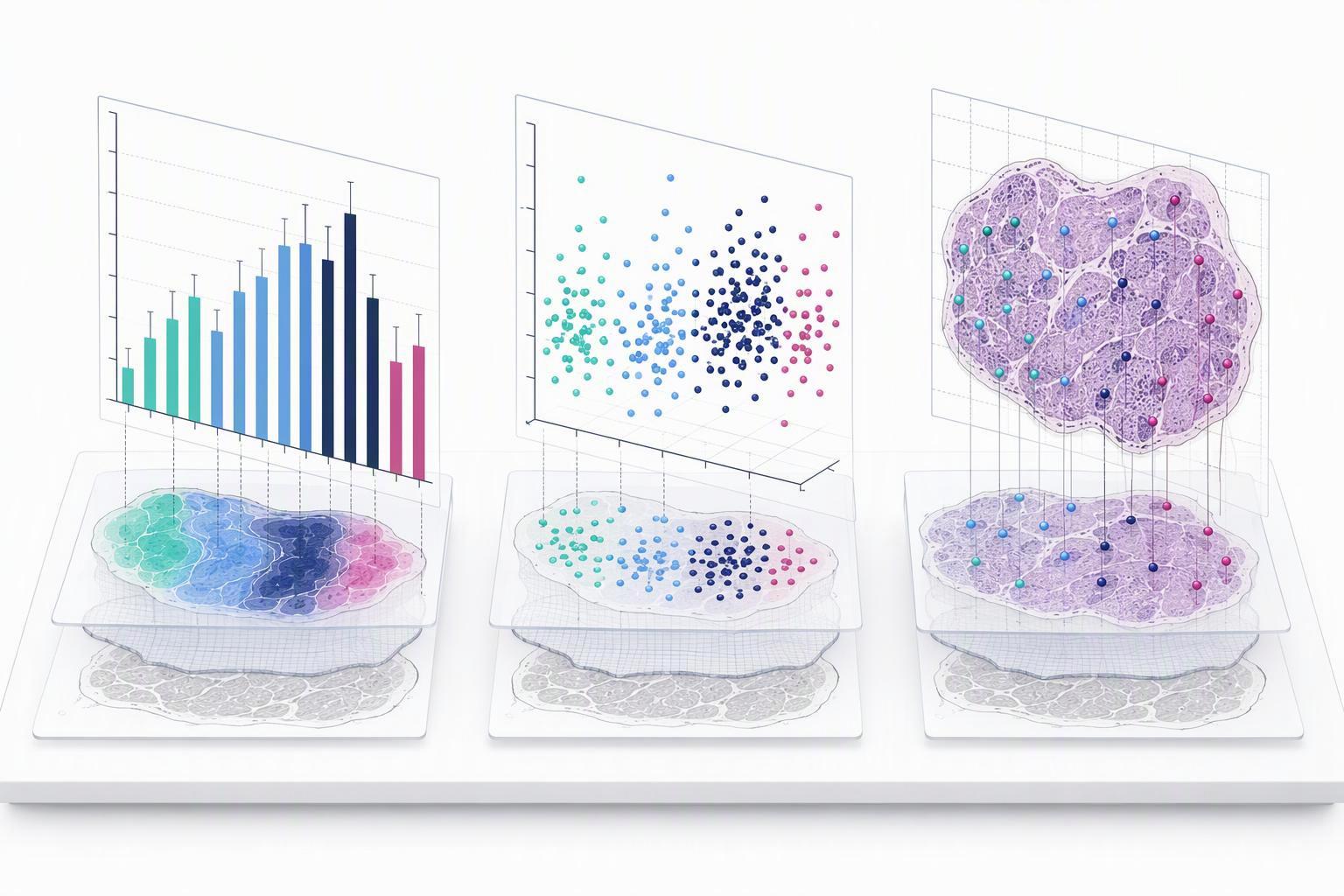
Veleprodaja sintetičkih polipeptida Solution Peptides Position is the variable that bulk sequencing destroys, and it is the variable a peptide probe has to survive. Spatial transcriptomics measures gene expression together with the physical coordinates of the RNA in intact tissue, so it resolves which cells or regions express a transcript, how close they sit to their neighbours, and patterns such as layers, niches and boundaries, whereas bulk RNA-seq averages across the tissue and dissociated single-cell methods lose position (spatial transcriptomics review, 2023). The founding method established the principle: histological sections placed on arrayed reverse-transcription primers carrying unique positional barcodes return quantitative expression plus the two-dimensional distribution of mRNAs, first shown in mouse brain and human breast cancer (the original spatial transcriptomics method, 2016).
The methods split into two classes with different failure modes. Imaging-based platforms use highly multiplexed single-molecule FISH to decode targeted genes without sequencing, while sequencing-based platforms use spatial barcodes on an array plus next-generation sequencing to restore position (the two technology classes in spatial transcriptomics, 2023). Neither class escapes the field’s central trade-off. A curated review of the literature dating back to 1987 documents the tension between resolution, throughput and sensitivity rather than a single best platform (the field’s own taxonomy of spatial methods, 2022).

The numbers make the trade-off concrete. In an independent benchmark that ran five commercial platforms on matched FFPE human tumour sections from six cancer types, Visium v1 and v2 use 55 µm spots, while Visium HD uses a contiguous 2 × 2 µm barcode grid over a 6.5 × 6.5 mm capture area, yielding roughly 11 million bins at native resolution, s 8 µm and 16 µm aggregations typically used instead (the matched-tissue platform benchmark, 2026). The same benchmark measured imaging panels at 377 genes for Xenium Multi-Tissue, 5,000 genes for Xenium Prime, i 1,000 genes for the CosMx Universal Cell Characterization panel, with Xenium scanning a continuous area up to 1.1 × 2.4 cm against discrete 0.5 × 0.5 mm fields of view on CosMx (the same benchmark’s measured panel sizes, 2026). For biomarker discovery, that geometry decides whether a rare invasive-margin population is resolvable at all, or whether it is averaged into a 55 µm spot alongside the stroma next to it.
Why does spatial biology for peptide design change target prioritization?
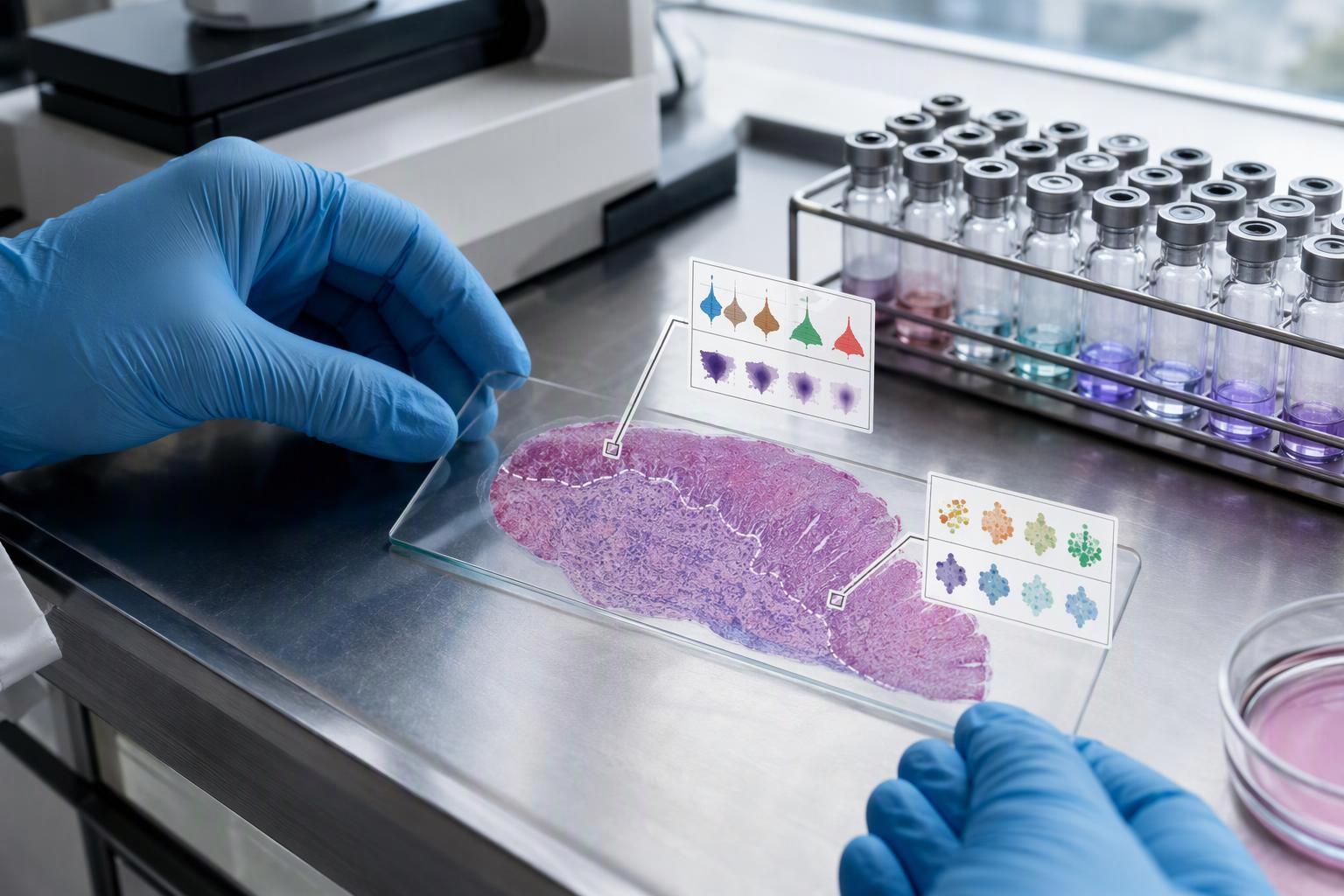
Prioritization changes because spatial data separates expression level from expression location, and a probe has to satisfy both. The first published rule for spatial analysis is to confirm that spatial information is essential before committing to the method at all, which is the same test a peptide program should apply to a candidate target (the ten published rules for spatial analysis, 2026). If a target’s value does not depend on where it sits, a bulk measurement answers the question more cheaply.

Use an analysis that respects the experimental unit, patient or animal replication, spatial dependence and multiple testing. These are important design checks; the cited sources do not establish that statistics is where most tissue-based programs fail.
This is the practical sense in which spatial biology for peptide design changes target prioritization: the ranking criterion shifts from abundance to accessible abundance in a defined compartment. A transcript enriched in a region the probe cannot reach ranks below a moderately expressed target on a surface the assay can sample. The evidence for that reframing is structural rather than statistical, and it comes from the discordance between RNA and protein measured in the same sections, which the next sections address.
How do localized expression and cell-state maps become peptide probe design criteria?
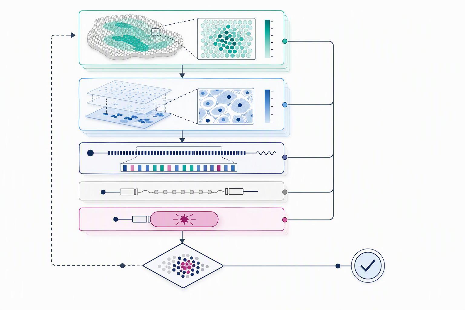
A probe has three parts, and spatial data constrains all three. The primary literature defines a probe as a targeting moiety, a linker and a reporter group, and states that protease-targeted probes need a peptide recognition sequence selective for the target protease plus a reporter (how peptide probes are constructed and selected, 2025). Localization data feeds directly into the first part: the recognition sequence has to match a target that is present in the compartment the probe will sample.
The same source draws a design distinction that spatial maps make actionable. Substrate-based probes amplify signal because one enzyme processes many probe molecules, while inhibitor-based probes bind covalently and stoichiometrically and give a direct readout of catalytic function (how peptide probes are constructed and selected, 2025). A cell-state map that shows a target enzyme restricted to Tvornica prilagođenih peptida a small region argues for the amplifying architecture, because the stoichiometric readout will be limited by how many target molecules the region contains. The stated in-vivo requirements are equally concrete: high target selectivity, meaning resistance to cleavage by off-target enzymes with similar specificity, fast activation kinetics with signal within minutes, circulatory stability and favourable pharmacokinetics (how peptide probes are constructed and selected, 2025).
Peptide chemistry itself has been tuned to imaging-grade precision, which sets an expectation for what a designed probe can achieve. A rationally redesigned E/K coiled-coil pair reached 4 nm localisation precision in Peptide-PAINT, protiv 3.6 nm for DNA-PAINT, while shortening the imager peptide to K18 reduced precision to 8.5 nm, and the pairs span a wide affinity range: E3/K3 at 21 amino acids with a dissociation constant near 20 nM, E22/K19 near 81 nM, and E22/K18 near 1.7 µM (how peptide pairs are tuned for imaging, 2020). Binding kinetics and affinity are design variables, not fixed properties of peptides. The same logic applies to a tissue probe: slijed, the linker and the reporter each move the detection threshold.
How does labeling strategy change what a spatial readout can show?
The label determines which molecular layer the readout reports, and the layers disagree. In the matched-tissue benchmark, spatial RNA–protein correlation averaged approximately 0.1 across all genes and samples, protiv grubo 0.7 in matched bulk tumour samples from CPTAC across seven cancer types (the matched-tissue platform benchmark, 2026). The spatial proteomics arm of that study used a 35-protein immuno-oncology panel on the same sections. A probe designed from transcript data alone is therefore designing against a measurement that predicts protein abundance poorly in situ, even where the bulk correlation looks reassuring.
Discordance is not uniform, which matters for panel design. The same study identified three distinct RNA–protein concordance patterns across genes rather than one discordance rate (the matched-tissue platform benchmark, 2026). Some targets will track between layers and some will not, so a program that validates a probe against transcript signal alone is testing the wrong layer for the discordant classes. The practical response is to choose the label that reports the layer the probe will encounter: an antibody-based readout for surface protein accessibility, a transcript readout for expression mapping, and a mass-tag readout where multiplexed protein targets are needed.
Label-free approaches remove the antibody question but impose their own constraints. Mass spectrometry imaging images thousands of molecules per experiment without labels or antibodies, and commercial MALDI-MSI reaches 5–10 µm lateral resolution on some instruments, with atmospheric-pressure MALDI using a novel optics system reaching 1.4 µm and transmission-mode MALDI with laser post-ionisation reaching 600 nm in brain tissue (label-free mass spectrometry imaging, 2024). The resolution comes at a cost that a probe designer must plan around: in MALDI-MSI, protein imaging at high spatial resolution loses ablated and detectable analyte as pixel size decreases, and matrix crystal size can severely limit achievable resolution (what limits MALDI spatial resolution, 2026). Higher resolution and lower sensitivity travel together.
How should assay format and controls be chosen from spatial data?
The format follows from what the spatial data says about cell density and compartment size, and the controls follow from the format. Imaging-based panels remain in the few-hundred-to-thousand-gene range because of optical crowding, rising binding-error risk with more hybridisation rounds, long imaging time and small imaging area, so a format that assumes full-transcriptome coverage is not available from either in-situ platform today (the two technology classes in spatial transcriptomics, 2023).
Assess segmentation and cell-assignment quality before interpreting localization. The cited dataset’s QC metrics describe that platform, panel and processing version; they do not establish that segmentation is the most frequently omitted control across laboratories.
Quality control and normalisation belong in the format decision, ne nakon toga. Low-quality spots or cells with extremely low gene counts or abnormally high mitochondrial fractions should be removed or flagged, and each spot should not be treated as equivalent to a single cell, because library-size normalisation of spot-based data confounds biology when spots contain multiple cells, Peptides Supplements which is why spatially aware methods such as SpaNorm were developed (the community workflow reference for spatial data processing, current release). A 55 µm spot contains several cells, so any per-spot intensity threshold in a peptide assay inherits that mixture.
How do you tell true target localization from artifact in tissue-based peptide results?
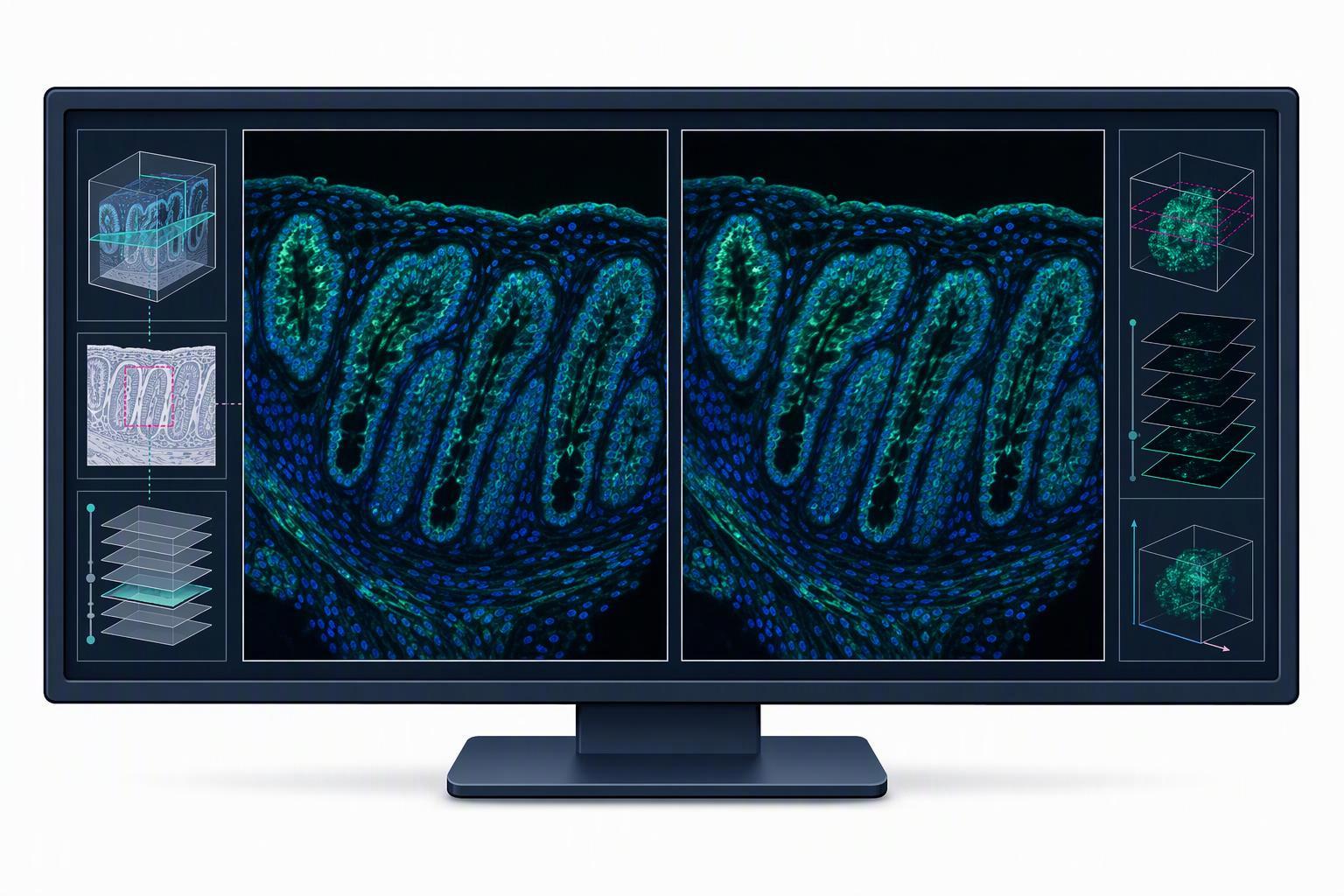
Artifact detection starts with the platform’s own geometry, because sampling area alone can shift the answer. In the benchmark, cell-composition estimates from the full Xenium scanned region differed from those in the CosMx-overlapping area in two of six samples, breast and colorectal, even though the two platforms agreed broadly when restricted to spatially matched regions (the matched-tissue platform benchmark, 2026). A target that appears enriched in one scan area and not another is a sampling artifact until proven otherwise. Wholesale Peptide Manufacturer
The benchmark also shows that platform differences are not always biological. Xenium consistently detected 5% do 30% more cells than CosMx, which the authors attribute to segmentation differences: Xenium uses DAPI staining to identify nuclei and computationally expands boundaries, while CosMx incorporates both nuclear and membrane staining (the matched-tissue platform benchmark, 2026). A cell-count difference of that size changes any per-cell normalisation, so a localization claim built on cell counts needs the segmentation method stated alongside it.
Two further checks separate signal from artifact. Prvi, the benchmark’s most directly comparable datasets were generated from serial sections processed together, while the Xenium 5,000-gene and segmented datasets were generated months later with updated reagents and are less directly comparable to the rest of the cohort, so cross-platform comparisons must carry that caveat (the matched-tissue platform benchmark, 2026). Drugi, alignment itself is a source of error: the benchmark aligned Xenium and CosMx coordinates by rotation angles and landmark shifts defined in image-editor software, whereas an open data framework now provides landmark-based alignment and coordinate transformations demonstrated on Xenium plus Visium breast cancer data (the open framework for sharing spatial datasets, 2024). Manual alignment steps are a documented artifact source, not a neutral preprocessing choice.
Where does spatially informed peptide work go next?
The data layer is becoming reusable infrastructure rather than a bespoke per-project effort, and that changes the economics of a probe program. The open framework for the field represents spatial data as five primitives, images, labels, points, shapes and tables, tracks the coordinate transformations and alignment steps applied to each dataset, and states its purpose as FAIR integration of multimodal spatial data and common coordinate frameworks (the open framework for sharing spatial datasets, 2024). A team that can query public spatial datasets before designing a probe spends its synthesis budget on targets that already have positional evidence.
The market figures should be read with their scopes attached, because they disagree by construction. The spatial biology market was estimated at approximately $1.14B in 2026 and forecast at approximately $2.46B by 2031, oko 17% annual growth, in a scope that includes multiplex immunofluorescence, digital spatial profiling, multiplex FISH and imaging mass cytometry instruments, reagents and services (the spatial biology market forecast, 2026). A differently scoped report puts the spatial omics market at USD 711.0M in 2024, USD 906.3M estimated for 2026 and USD 1,696.5M by 2030 na a 16.3% CAGR, with consumables at 56.2% od 2024 revenue and North America at 57.4% (the spatial omics market data, 2025). The two are not the same measurement, and blending them into one figure would misstate both. What they agree on is direction and the consumables-heavy mix.
The honest limit on all of this is that a peptide probe’s detection threshold in tissue remains poorly specified in the public literature. No source read here gives a numeric detection limit in fmol or amol, or a receptor-density threshold, for a peptide probe in a tissue assay, and the MALDI review states the resolution-versus-analyte trade-off without quantifying a floor (what limits MALDI spatial resolution, 2026). Sensitivity falls with pixel size, and penetration and background depend on label chemistry, but a team cannot yet design against a published number. That gap is the most useful thing a spatial dataset can help close internally, by measuring the threshold on the team’s own tissue before committing to a sequence.
Često postavljana pitanja
Can spatial transcriptomics replace protein-level target validation?
Ne. RNA abundance is not a measurement of surface protein, so a transcript map cannot confirm that a target is present, folded and reachable on the cell surface. The gap is large enough to matter: in the matched-tissue platform benchmark, spatial RNA and protein signals tracked each other at roughly 0.1, while the same analytes in bulk tumour material agreed at about 0.7 (the matched-tissue platform benchmark, 2026). Treat a transcript result as a reason to run the protein assay, ne kao zamjena za to. Peptide Supplements
Does high HPLC purity mean a peptide probe will be tissue-specific?
HPLC area purity is a detector- and method-dependent peak-area ratio, not necessarily the target’s mass fraction. It does not establish binding selectivity. Test probe identity, target recognition, tissue accessibility and relevant off-target binding using appropriate controls.
How many biological replicates does a tissue-based peptide study need?
Count replicates at the patient, animal or sample level, never at the spot or cell level. Spatially autocorrelated spots are not independent observations, and treating them as such inflates significance, which is the pseudoreplication problem the published rules for spatial analysis call out directly (the ten published rules for spatial analysis, 2026). The number itself depends on effect size and tissue heterogeneity, so power it against the biological unit you are actually sampling.
Can Visium and Xenium datasets be compared directly?
Only when the comparison respects how the data were generated. Serial sections processed together in the same run are comparable; datasets produced months apart, on different reagent versions or with updated panels, are not, because the measured panel and chemistry differ. The benchmark’s own caveats carry this limit, and its panel sizes illustrate the spread: 377 genes for Xenium Multi-Tissue, 5,000 for Xenium Prime and 1,000 for the CosMx Universal Cell Characterization panel (the matched-tissue platform benchmark, 2026). Match platform, panel and processing batch before you put two datasets on one axis.
What should I do when spatial RNA and protein disagree?
Treat the disagreement as the expected case rather than an anomaly, and resolve it at the protein level. Discordance is the baseline behaviour of these two layers in tissue, not a sign that one dataset is broken, so the decision that follows is which measurement answers your question. If the question is whether a probe can reach its target in a given compartment, the protein readout decides it, and the transcript map supplies the positional context around that answer.
Zaključak
Spatial data changes peptide design decisions at the point where abundance stops being the only criterion. The matched-tissue benchmark’s approximately 0.1 spatial RNA–protein correlation against roughly 0.7 in bulk is the clearest evidence that a probe designed from transcript rankings alone is designing against the wrong layer (the matched-tissue platform benchmark, 2026). The controls that make a tissue result defensible are equally specific: count replicates at the sample level, state the segmentation method, and treat sampling area as a variable that can move the answer.
The next step for a team starting this work is to read the method before committing the sequence. The ten published rules for spatial analysis are a short, practical checklist covering platform choice, workflow structure, segmentation tools and the requirement to acknowledge technical limitations, and they are worth an hour before the first section is cut (the ten published rules for spatial analysis, 2026). Read them alongside the matched-tissue benchmark, and decide which compartment your probe has to reach before deciding what it should recognize.