
The conversion is a redesign, not an add-on
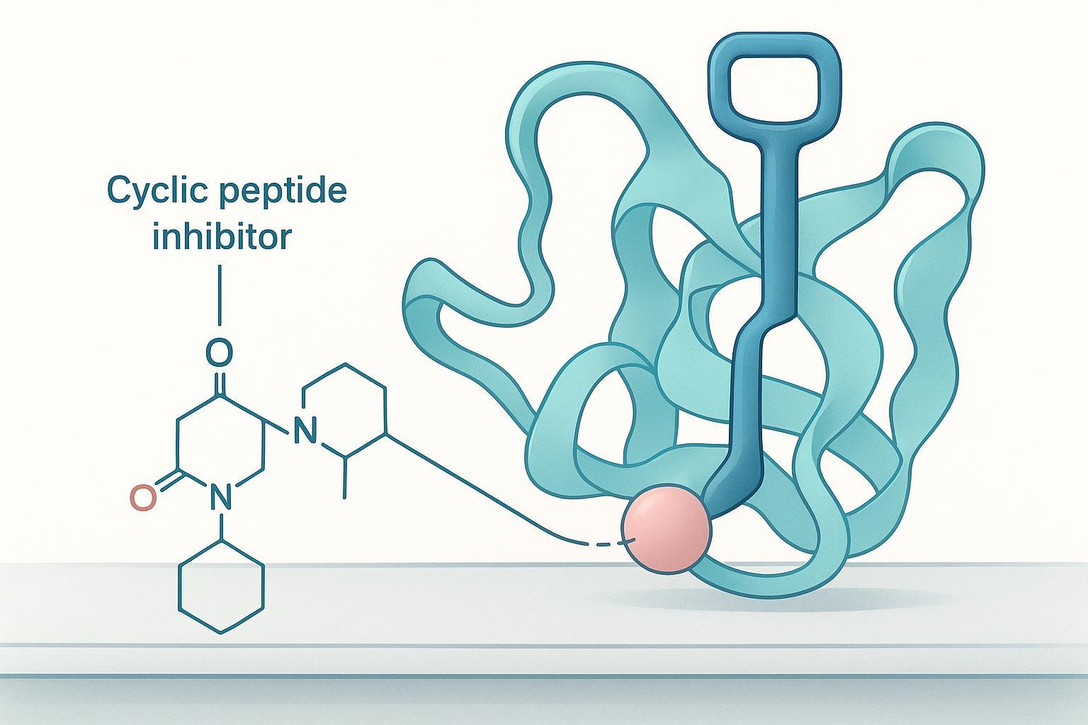
A cyclic peptide that inhibits HIF-1α by occupying the PAS-B pocket is a key cut to fit one lock. Turning it into a degrader means welding a handle onto that key: a linker plus a VHL-recruiting element, positioned so the ring still seats in the pocket while the new arm reaches out to recruit the ligase. The teeth cannot change. Everything else can.
That distinction drives the whole project. In the anchor case, the linker and VHL ligand were built in as a custom bifunctional amino acid and incorporated directly during solid-phase peptide synthesis, rather than being attached after cyclization (JACK, Cyclic Peptide PROTACs Restore VHL-Mediated HIF-1α Degradation in Hypoxia, 2026). The practical consequence is that the attachment point becomes a design choice fixed at chain-assembly time, not a variable you tune later at the bench.
Every downstream decision follows from that commitment: which gradient resolves the conjugate, which conformational check confirms the ring survived, and which assay can distinguish degradation from mere occupancy.

Which exit vector should carry the linker?
Conjugation-site selection is the first decision that constrains every later one, and the rule is simple to state: choose the position least likely to perturb binding and most likely to be solvent-exposed (Current strategies for the design of PROTAC linkers, 2020). When a co-crystal structure of your cyclic peptide bound to the target is available, that rule is answerable by inspection. When it is not, inspection cannot decide the question, and pretending otherwise is how programs lose months.
Dịch vụ The attachment point is also not a derivatization step you bolt on after the peptide is optimized. It is a chain-assembly design choice, fixed before synthesis begins, as it was in the anchor case. Changing your mind later means resynthesizing the peptide, not editing a side chain.
The empirical ranking rule: rank candidate vectors by what each one preserves, in this order: target affinity, then ternary-complex behaviour, then degradation. A narrow SAR at a position that looked solvent-exposed is a warning, not noise. That position is conformationally load-bearing, and the ranking has to be redone.
Installing the linker and the E3 element
The route decision comes first, because on-resin and solution conjugation fail in different places. On-resin installation of a bifunctional amino acid builds the linker into the chain itself, so the protecting-group scheme has to keep the linker’s orthogonal handle intact through Cửa hàng every subsequent coupling and the cyclization step. Solution conjugation after cyclization is faster to iterate, but it exposes the free peptide to activation chemistry twice and leaves you purifying a mixture of positional isomers if the reactive residue is not unique.
The Southampton work shows what that iteration buys. The series that began with cyclo-CLLFVY moved to a second-generation warhead, cyclo-CLLF(CF3)IF(Br), carrying longer unnatural amino acid linkers built into the sequence rather than appended afterwards (McDermott et al., 2025). Building the linker into the chain is what makes linker length a design variable instead of a purification problem.
That matters because the main threat to peptide purity and folding on this route is epimerization, and it is mechanistically avoidable. Activating an amino acid forms a planar oxazolone that is temporarily achiral at the α-carbon, so reprotonation can return either the L- or the D-configuration. Risk climbs with extended activation, highly reactive coupling reagents, and accumulated activated intermediate, and residues such as phenylglycine, cysteine and histidine are especially prone (Bachem; Epimerisation in Peptide Synthesis, 2023).
The magnitude is route-dependent, and one vendor’s feasibility work illustrates the spread rather than setting a benchmark: fragment-route conjugation produced roughly 13% epimer formation and was discontinued, while the alternative route showed 2–3% epimerization rising to about 5% after purification, and linear SPPS gave the highest yield with no epimerization detected (Bachem). Read that as a signal about which route classes carry the risk, not as a rate you can expect on your own sequence. Về
What RP-HPLC purity data can and cannot tell you
A single main-peak percentage at 214 nm confirms neither correct conjugation nor correct folding. It tells you that the UV-absorbing species in your sample elute as one band on one gradient. That is a useful fact, and it is a smaller fact than most programs treat it as.
For a larger, more hydrophobic conjugate, the method resolves less than it did for the parent cyclic peptide. The linker and E3 element add hydrophobic surface area, the retention time shifts, and species that were baseline-separated on the parent gradient start to co-elute. A published disulfide-cyclized peptide degrader program reports its preparative practice plainly: one RP-HPLC step on a 0.1% TFA water/acetonitrile gradient running 26–43% over 40 minutes at 10 mL/min, detection at 220 bước sóng, oxidation monitored by mass spectrometry alongside HPLC, and a final HPLC quality check before lyophilization (Frontiers in Immunology, 2023). Note what that paper does not state: any purity threshold. The number is a process output, không phải là một đặc điểm kỹ thuật.
Suppliers commonly specify ≥95% RP-HPLC purity as the floor for biological evaluation, ≥98% for quantitative binding and dose-response work, and ≥99% for reference-standard work, with purity defined as main-peak area over total peak area by UV detection at 214–220 nm (GenScript; Proteomics sáng tạo, 2025). Treat those bands as purchasing convention rather than a measured finding about your molecule. They describe what a vendor will certify, not what your conjugate needs.
The gap matters because peptide purity and folding are different questions, and RP-HPLC answers only the first. A co-eluting epimer, a misfolded disulfide isomer, or an unreacted parent that happens to run at the same retention time all present as a clean single peak. Mass identity, stereochemistry, and solution conformation each require an orthogonal method.
Read the matrix as a coverage check, không phải là một thứ hạng. RP-HPLC covers hydrophobicity and purity. LC-MS and HRMS cover mass identity, which is how you confirm the linker actually installed. Chiral LC covers stereochemistry, which is how you catch epimerization at the conjugation site. NMR and CD cover solution conformation, which is how you distinguish a folded conjugate from an unfolded one. No single row covers more than one column, and a program that reports only the first row has evidence for exactly one of four questions.
The practical consequence for conjugation-site selection: if your analytical package is one RP-HPLC trace, you cannot yet tell whether the exit vector you chose produced the conjugate you designed. Add the orthogonal methods before you commit the route, not after a binding assay comes back flat.
Folding and conformational verification
Tổng hợp peptit Cyclization constrains the backbone, but it does not guarantee that the bioactive conformation survives linker addition. That distinction is why conformational evidence has to be orthogonal to purity evidence: each method class answers a question the others cannot.
Chiral LC resolves stereochemistry, so it tells you whether the conjugate racemized at the residues you care about. CD reports on solution secondary structure, and NMR reports on the specific contacts that define the bound fold. None of these substitutes for another, and none of them substitutes for a functional readout. The same principle governs the assay stage: a ternary-complex signal alone is insufficient, and cellular DC50/Dmax values alone are phenotypic rather than mechanistic (Methods to Study the Molecular Mechanism and Drive the Design of Degraders, đã lấy lại 2026-05-14).
Chìa khóa mang đi: High HPLC purity is not correct conjugate. A peak that is clean and mass-confirmed can still carry the wrong fold into the assay stage.
For your data package, that gap is the risk to close. A conjugate that passes purity and mass identity but was never conformationally checked carries an unquantified risk forward, and the assay team inherits it. Treat peptide purity and folding as two separate evidence lines, each with its own method and its own acceptance criterion, before the material is released to biology.
The functional assay evidence chain
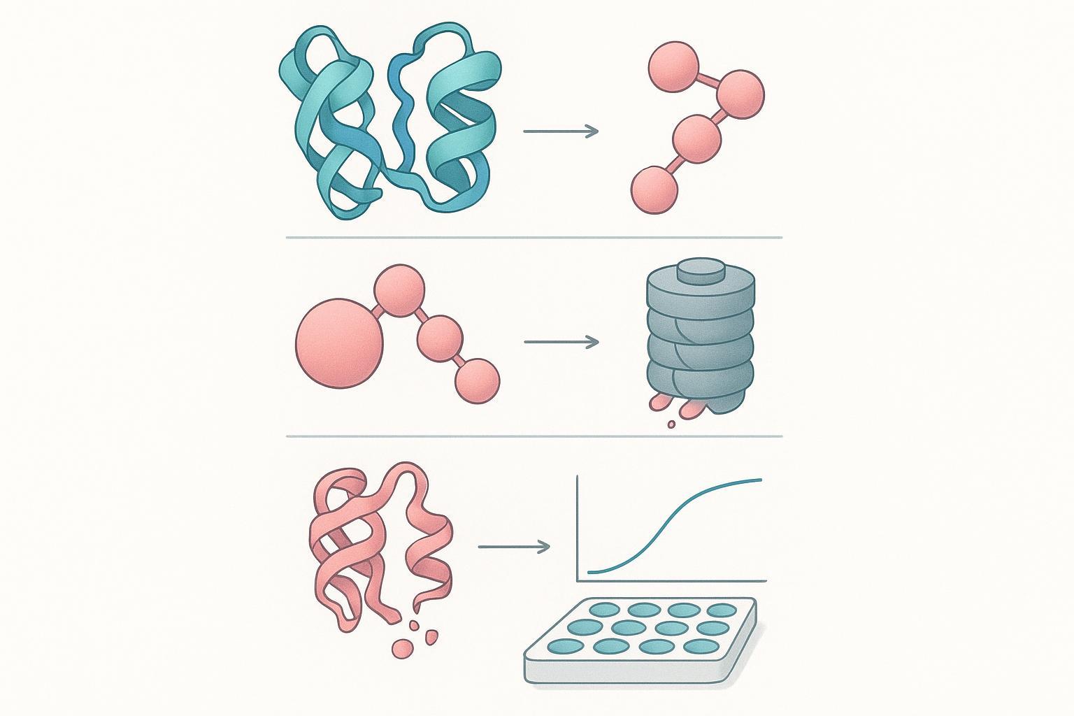
A degradation claim is a mechanism claim, and no single assay carries it. The evidence chain has four links, and a reviewer will look for all four: ternary complex formation, target ubiquitination, proteasomal dependence, and a quantitative potency and ceiling readout. If your package stops after the first link, you have shown binding, not degradation.
Work down the chain against your own data. Ternary complex: a co-crystal or a biophysical assay showing target, PROTAC and E3 ligase in one assembly. phổ biến: a direct or indirect readout that the target itself acquires ubiquitin. Proteasomal dependence: rescue by a proteasome inhibitor. Potency and ceiling: DC50 and Dmax from the same dose-response series.
The third link is the one most often skipped, and it is cheap to close. In the cyclic peptide work, PD-L1 loss at 2.5 µM after 4 hours was blocked by MG132, which is what separates degradation from a downstream transcriptional effect (Frontiers in Immunology, 2023). Target engagement can be shown the same way: cellular thermal shift assays moved DHHC3 from 49 °C in untreated cells to 55 °C in peptide-treated cells, evidence that the compound reaches the protein before any degradation is measured (Frontiers in Immunology, 2023).
That chart is the strongest quantified argument in this article for treating the E3 element as a design variable rather than an afterthought. Swapping CRBN for VHL, with the same warhead chemistry, moved DC50 from 0.103 µM to 1.92 µM, and the IAP arm to 7.530 µM (Cell Death & Disease, 2024). Nearly two orders of magnitude separate the best and worst arm in the same cellular assay.
Now the gap. The same series reports DC50 values but no Dmax, so potency is comparable across arms while the ceiling of degradation is not (Cell Death & Disease, 2024). A compound can be potent and still leave most of the target standing. When you hand a package from chemistry to biology, ask for the full dose-response curve, not the single number: functional assay support means the curve, the rescue control, and the engagement assay together, because that is what a reviewer will ask for on HIF-1α degradation or any other target you take forward.
The oxygen-state limit on the anchor case
The anchor cyclic peptide PROTAC restores VHL-mediated HIF-1α degradation robustly at 3% O₂, and shows no net degradation at 1% O₂. That is not a potency failure. Under the reported conditions the degrader stays engagement- and degradation-competent at 1% O₂, but the target’s resynthesis outpaces clearance, so the steady-state pool never falls (JACK, Cyclic Peptide PROTACs Restore VHL-Mediated HIF-1α Degradation in Hypoxia, 2026).
Read that as a design rule rather than a footnote: a degradation readout is only interpretable against the resynthesis rate of the target in the same cellular context. A DC50 measured at one oxygen tension does not transfer to another, because the number you measured is a net balance, not a clearance constant.
Before you compare your conjugate against a published DC50, confirm the oxygen tension, the cell line, and the resynthesis behavior of the target under that same condition.
Common misconceptions that cost programs time
Four decision errors account for most of the lost time in a cyclic peptide PROTAC design program. Each one substitutes a cheaper measurement for the question the program actually needs answered.
High HPLC purity is not a correct conjugate. A single sharp peak at 95% tells you the material is homogeneous by one detection method. It does not tell you the linker sits on the intended residue, that the stereochemistry survived conjugation, or that the E3 ligand is intact. The fix is orthogonal characterization: LC-MS for mass confirmation, and a second chromatographic or spectroscopic method that separates the conjugate from its positional isomers.
Retained target binding is not degradation achieved. A binding assay reports occupancy. Degradation requires ubiquitination, proteasomal engagement and target loss, none of which a binding readout detects. The fix is to treat binding as a gate, not an endpoint, and to require target protein quantification before claiming degradation.
A ternary complex in a biophysical assay is not cellular degradation. Ternary complex formation is necessary but not sufficient; a stable complex can still fail to ubiquitylate. The fix is to carry the claim into cells and measure target protein levels directly.
A DC50 without a Dmax is a partial potency picture. Potency and extent are separate properties, and a compound that degrades 40% of the target at saturation is a different proposition from one that clears 90%. The fix is to report both, luôn luôn, from the same dose-response series.
A decision framework for the conversion
Run these five checks in order against your own program. Each one has a failure mode attached, and each one is cheaper to fix before the next.
-
Rank candidate exit vectors empirically, not by inspection. Express the same cyclic inhibitor with the linker at two or three different positions, run the conjugate series through the target-binding assay, and let the rank order pick the winner. Chế độ lỗi: choosing the vector that looks most accessible on the model and discovering after scale-up that binding is gone.
-
Decide the route before you commit the linker. On-resin versus solution conjugation determines how much epimerization risk you carry, so treat it as a route decision rather than a late optimization. Chế độ lỗi: a conjugate that purifies cleanly but has lost the stereochemistry the binding data depended on.
-
Purify, then characterize orthogonally. RP-HPLC purity and mass confirmation answer different questions, and neither one reports on conformation. Chế độ lỗi: a single-method release criterion that passes a misfolded batch.
-
Verify folding before you spend assay budget. Circular dichroism, NMR, or a structure-sensitive binding readout confirms the conjugate still occupies the intended conformation.
-
Build the assay chain before claiming degradation. Ternary complex formation, target engagement, Peptide tổng hợp and a degradation readout are three separate claims, and a binary binding result supports only the first.
Chìa khóa mang đi: Conformational verification is the item programs skip most often. Purity data and mass confirmation cannot tell you whether the conjugate folded correctly, so a batch can pass every analytical gate and still fail functionally.
The sequence matters more than any single step: a program that ranks vectors first and verifies conformation before assay work avoids re-synthesizing a conjugate it has already characterized.
Các bước tiếp theo
If a cyclic peptide PROTAC design program is on your roadmap, the useful next move is a scoping conversation rather than a purchase decision. Bring three things: your anchor sequence and any structural data, the exit vector you have chosen or the candidates you are weighing, and the assay readouts your team will accept as evidence of degradation. Those three inputs determine whether the conversion is a straightforward linker installation or a redesign that needs new chemistry.
For teams that need a synthesis and characterization partner, MOL Changes states capabilities across solid-phase and microbial fermentation synthesis, controlled-environment purification, HPLC/MS and sterility QC, and mg-to-kg scale. That range matters most when a conjugate program moves from analytical-scale material to the quantities a functional assay series requires. Sản xuất peptit
Chìa khóa mang đi: The constructs described here are research reagents. Any clinical translation requires independent validation, and nothing in this guide substitutes for your own analytical and biological confirmation.
Tiết lộ: the author’s organization provides peptide synthesis and characterization services of the kind discussed in this article.
Discuss a custom conjugate program. Share your anchor sequence, target exit vector and required assay endpoints, and request the analytical documentation package (purity method, độ dốc, MS confirmation and folding data) alongside a scoping call. Start a program discussion.
Frequently asked questions
What do I do if there is no co-crystal structure of my cyclic peptide bound to the target?
Rank candidate attachment positions empirically instead of waiting for a structure. Express the peptide with the linker at each candidate residue, then measure in sequence: preserved target affinity, ternary-complex formation with the E3 ligase, and cellular degradation. A position that tolerates substitution keeps its affinity; one that does not is disqualified. Treat narrow structure-activity relationships at a candidate position as a stop signal, because the exit vector is probably buried in the binding interface.
Is a longer linker always better for a cyclic peptide PROTAC?
KHÔNG. Linker length and composition change ternary-complex geometry, and that geometry drives potency in both directions. The frequently quoted 5-15 atom window comes from secondary recaps whose primary pages could not be read in this run, so treat any specific range as a starting hypothesis to test across a small series, not a specification to design against.
How do I distinguish epimerization from a folding problem on my analytical data?
Use orthogonal methods, because a single RP-HPLC main peak cannot separate the two failure modes. Chiral LC or NMR resolves stereochemistry at the affected residue; circular dichroism and NMR report on solution conformation. Epimerization is a known hazard of the conjugation chemistry rather than a vendor talking point, so when peptide purity and folding both look acceptable and activity still drops, run the stereochemical check before redesigning the linker.
What is the minimum assay set to claim degradation rather than inhibition?
Four measurements: ternary-complex formation, target ubiquitination, proteasomal dependence confirmed by MG132-type rescue, and quantitative DC50 and Dmax from dose-response curves. Ternary complex alone is insufficient, since binding without ubiquitination transfer proves nothing about degradation. DC50 and Dmax alone are phenotypic: they show the target disappeared without showing the proteasome caused it. Report the set together, and state the cell line and time point for each.